1 Basic knowledge of optical microscopy – propagation of light
(1) Reflection
When light propagates to different materials, it changes the direction of propagation at the interface and returns to the original material. This phenomenon is called the reflection of light. The reflected ray is on the same plane as the incident ray and the normal; the reflected ray and the incident ray are separated on both sides of the normal; the reflection angle is equal to the incident angle. Light is reversible. In the phenomenon of light reflection, the optical path is reversible.
(2) Refraction
When light is incident obliquely from one transparent medium into another transparent medium, the direction of propagation generally changes. This phenomenon is called refraction of light. The speed of reflected light is the same as that of incident light, while the speed of refracted light is different from that of incident light.
(3) Interference
Interference is a phenomenon in which two or more waves overlap in space and form a new waveform. When two columns of light waves with the same frequency meet in the air, they are superimposed. They are always strengthened in some areas and weakened in other areas. The phenomenon of light and dark stripes or colorful stripes is called light interference. Only coherent light sources with the same frequency, constant phase difference, and consistent vibration direction of the two columns of light waves can produce light interference.
(4) Diffraction
When light encounters an obstacle or a small hole during propagation, the light will deviate from the straight path and propagate behind the obstacle. Only when the size of the small hole or obstacle is similar to or smaller than the wavelength of the light wave can significant diffraction occur.
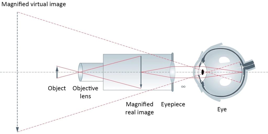
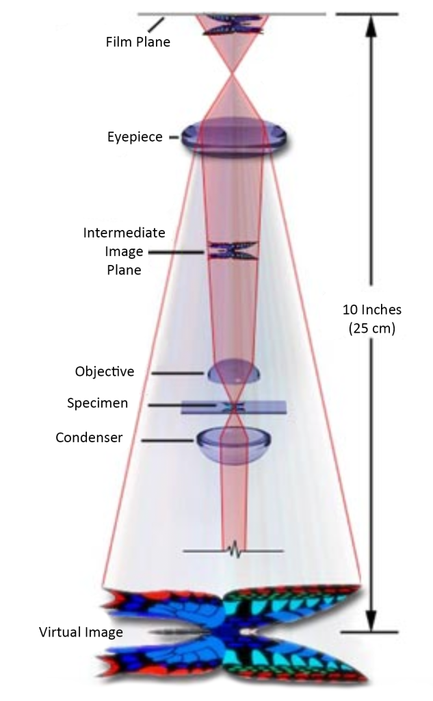
2 Optical principles of optical microscopes
An optical microscope is an optical instrument that uses optical principles to magnify and image tiny objects that cannot be distinguished by the human eye so that people can extract detailed structural information. Due to the differences in samples, using the same observation method to observe different samples often cannot achieve ideal imaging. Therefore, by choosing different observation methods for different samples, the imaging problem can be easily solved.
3 Seven imaging methods of optical microscopes
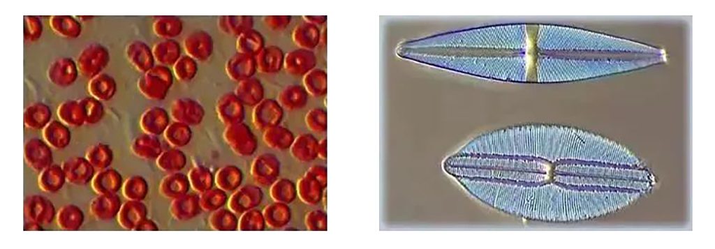
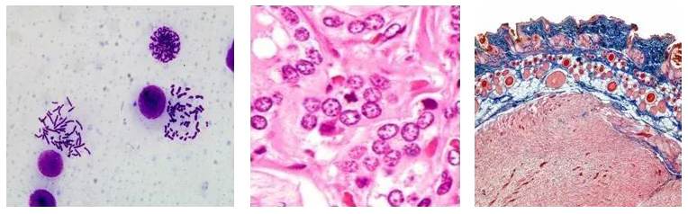
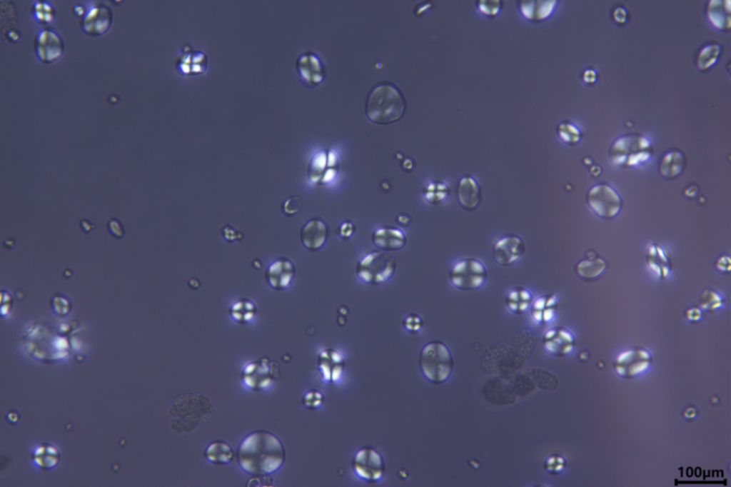
(1) Bright-field-BF
Brightfield microscopy is the simplest of all light microscopy illumination techniques. Sample illumination is transmitted (i.e. illuminated from below and viewed from above) white light, and contrast in the sample is caused by the attenuation of transmitted light in dense areas of the sample. Brightfield microscopy is the simplest of a series of techniques used to illuminate specimens in light microscopy. The typical pattern of brightfield microscopy images is a dark sample on a bright background, hence the name.
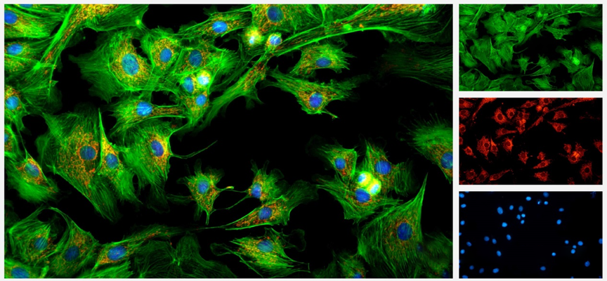
(2) Fluorescence Field-FL
Fluorescence observation is the most commonly used observation mode among friends. So what is fluorescence? That is, the electrons in the substance absorb light energy and change from a low-energy state to a high-energy state, and then the light released when returning to the low-energy state is non-temperature radiation – luminescence. In short, it means that matter absorbs short-wave light and emits long-wave light. Its characteristics are that it must absorb light, that is, there must be an excitation light source; the fluorescence wavelength > the excitation wavelength; and the intensity of the fluorescence must be less than the intensity of the excitation light. The intensity of fluorescence mainly depends on three major categories: concentration of the test substance, fluorescence efficiency and excitation light intensity. It has unique advantages: high specificity – targeted labeling; little stimulation to cells – in vivo staining; capable of multiple staining – co-localization. It is mostly used for positioning and qualitative and quantitative research.
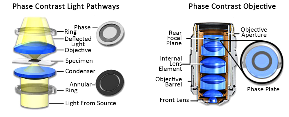
(3) Phase contrast-Ph
Phase contrast observation is also one of the commonly used methods of microscopic observation, which requires special phase contrast objectives. The principle is to use the optical path (refractive index x thickness) difference of the object to be inspected for microscopic inspection, that is, using the interference phenomenon to change the phase difference into an amplitude difference that can be distinguished by the human eye. The phase difference of light cannot be felt by the naked eye, but the phase contrast microscope can use its special device – annular diaphragm and phase plate to use the interference phenomenon of light to convert the phase difference of light into an amplitude difference (light and dark) that can be detected by the human eye. Difference), so that the original transparent object shows an obvious difference between light and dark, and the contrast is enhanced, allowing us to observe some subtle structures more clearly.
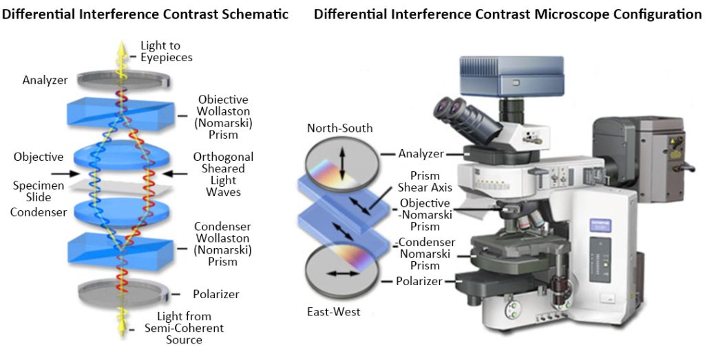
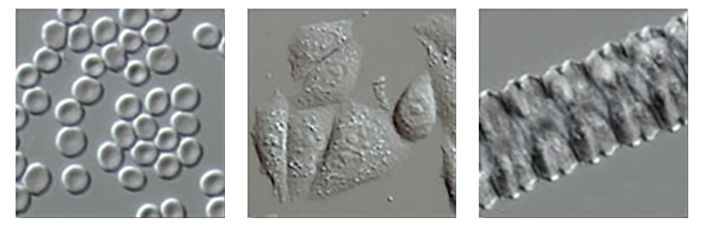
(4) Differential Interference Contrast-DIC
The physical principle of differential interference is completely different from that of phase contrast microscopy, and the technical design is much more complex. DIC uses polarized light. The transmission DIC microscope has four special optical components: polarizer, DIC prism, DIC slider and analyzer. DIC microscopy makes the structure of cells, especially some larger organelles, such as nuclei, mitochondria, etc., have a particularly strong three-dimensional sense, making it suitable for micromanipulation. At present, microscopic operations such as gene injection, nuclear transplantation, transgene, etc. are often performed under this microscope.
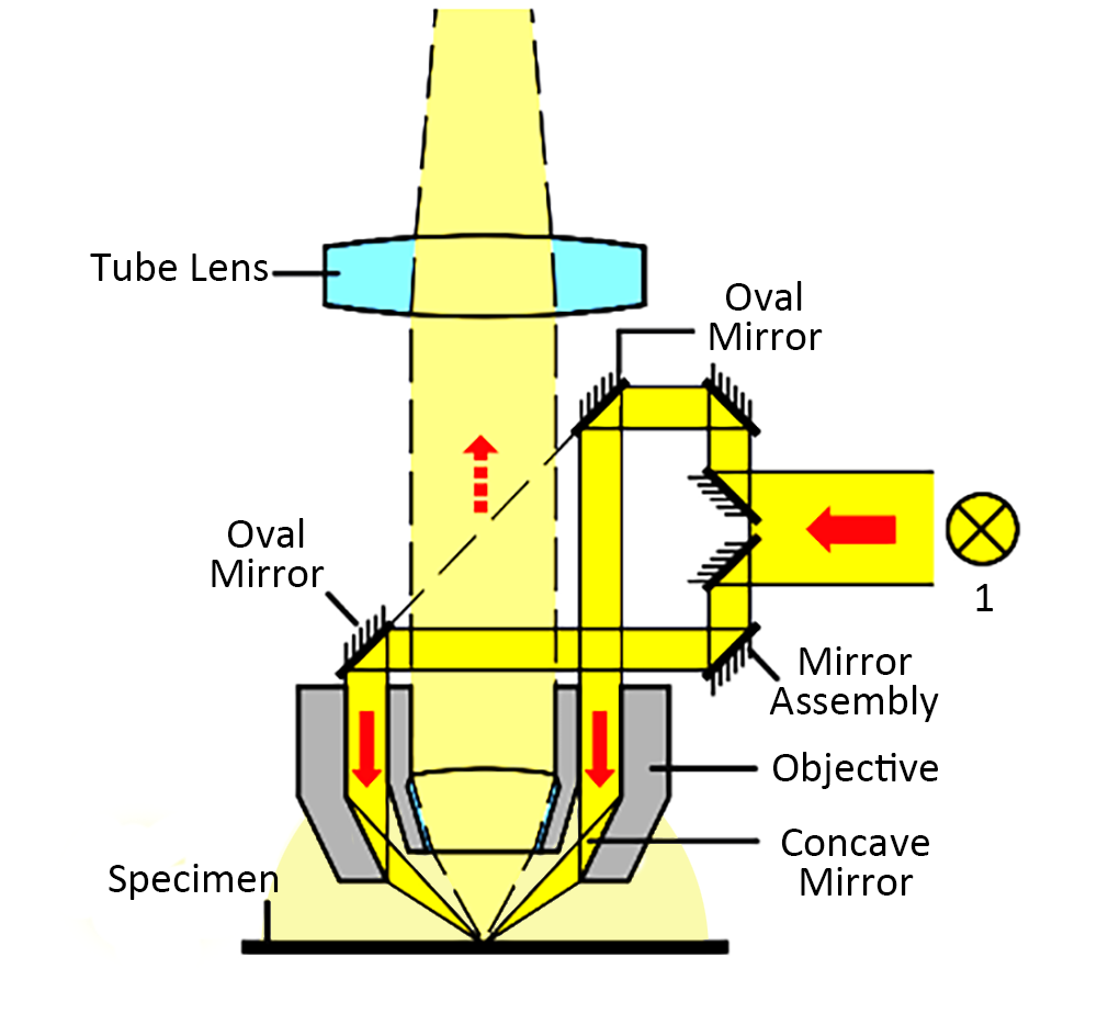
(5) Dark Field
Dark-field imaging corresponds to bright-field imaging. As the name suggests, people do not observe the illumination light directly, but the light slants to the surface of the specimen. According to the Tyndall phenomenon, the particles are Oblique light is reflected or diffracted, which increases the visibility of the human eye, so that we can observe extremely small objects. So how to achieve dark field imaging? Looking through the microscope cross-section, we can see that the condenser aims a cone of light at the specimen at an altitude angle so that some of the light does not directly enter the objective lens. Light passing through the specimen is diffracted, reflected, and refracted by optical discontinuities (such as cell membranes, cell nuclei, and internal organelles), reenters the objective lens, and the specimen can then be seen as a bright body on a black background.

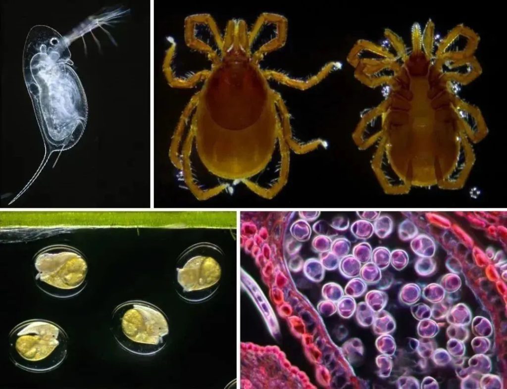
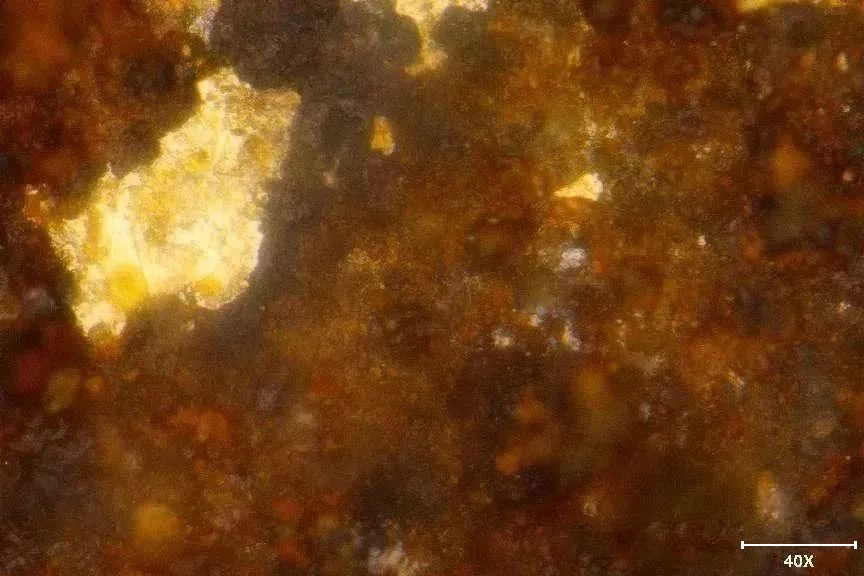
The reflected light darkfield can reflect the ultra-tiny structure of the sample surface, and structures as small as 0.004um can be observed. However, due to the scattering or diffraction of light by small structures, accurate measurements cannot be carried out in the dark field. Therefore, the dark field is only suitable for observation and used to locate some ultra-small structures and defects. Of course, the reflected light darkfield also has another small use, which can obtain the color of transparent or translucent materials, such as the green oil of PCB boards, the color of rust, etc.
(6) Polarized Light
The polarizing microscope is an essential instrument for studying and identifying birefringent substances using the polarization characteristics of light. It can perform single polarization observation, orthogonal polarization observation, and colonoscopic observation. A method of microscopic examination by changing the polarization of ordinary light to identify whether a substance is monorefractive (isotropic) or birefringent (anisotropic). Birefringence is a basic characteristic of crystals. Therefore, polarizing microscopes are widely used in minerals, chemistry, and other fields. In humans and zoology, polarized light microscopy is often used to identify bones, teeth, cholesterol, nerve fibers, tumor cells, striated muscle, and hair.

(7) Huffman Modulation Contrast (HMC)
Hoffman-modulated phase contrast uses oblique light irradiation to convert phase gradients into changes in light intensity, which can be used to observe unstained samples and living cells. Its system usually consists of a condenser with a slit and a rotatable polarizer in the condenser. It is suitable for high-resolution microscopic observation of phase objects, birefringent objects, stained samples, columnar objects, and objects with surface and subsurface features. It can conduct dynamic analysis of cell tissues, transparent liquid tissues, and cultured tissues in culture dishes. Microscopic observation and scientific analysis.