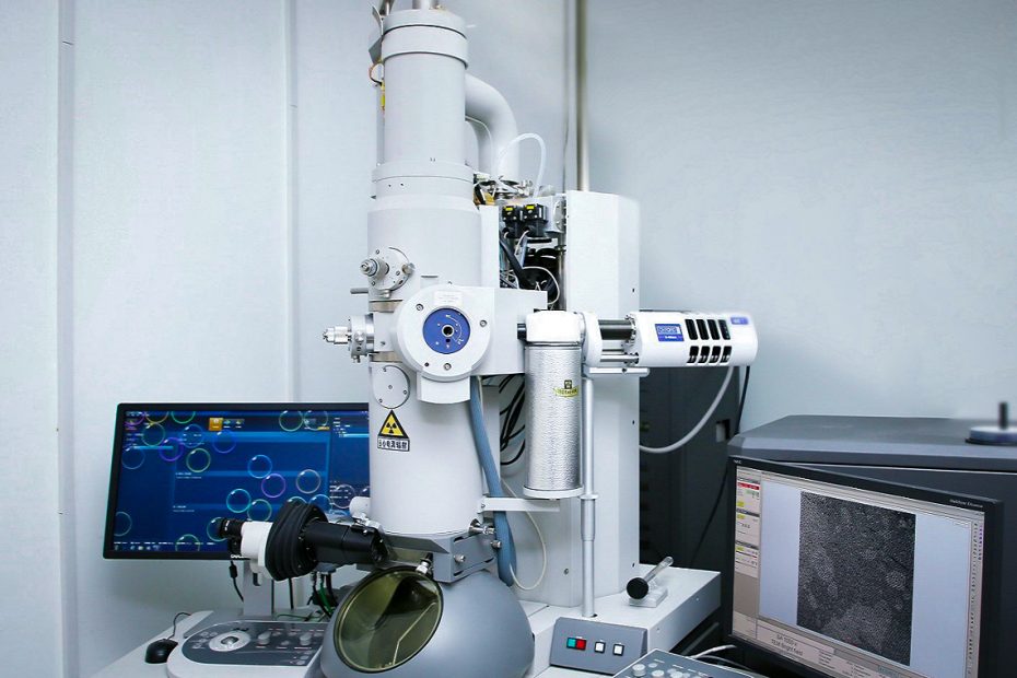
Scanning electron microscopy (SEM) is widely used for nanofabrication characterization, metrology, and process control. I believe that everyone will encounter vibration and drift problems when doing SEM analysis, resulting in the inability to obtain high-definition images and affecting the accuracy of the analysis results.
Reasons for electron beam deflection or drift in a scanning electron microscope may include the following:
Environmental factors: Factors such as temperature, humidity, vibration, etc. may affect the accuracy of the scanning electron microscope, causing the electron beam to drift or offset.
Uneven sample surface: An uneven or rough sample surface may affect the scanning quality of the electron beam, causing the electron beam to drift or shift.
Electron Gun Deviation: Electron gun deviation can cause the electron beam to be oriented incorrectly, causing the electron beam to drift or shift.
Problems with the electron beam itself: Problems such as poor focus of the electron beam and uneven energy distribution of the electron beam may cause the electron beam to drift or shift.
This article discusses measurement uncertainties caused by vibration and drift, along with some possible solutions.
During the SEM image acquisition process, the equipment may be adversely affected by the surrounding environment. Environmental machinery and noise will significantly affect electron microscope performance. The SEM’s column is directly coupled to the sample stage, so any external vibrations transmitted to the column through the frame and isolation system can ultimately be transmitted to the sample. Transmitted vibrations can produce unwanted artifacts in recorded images.
Additionally, sample stage movement and friction in its components can lead to drifting, and uncontrolled motion. Increased temperature and interaction of the electron beam with the electromagnetic field are also causes of drift. All of these can damage SEM image quality, as drift can manifest as a degree of severe image distortion, and elongation and vibration often manifest as jagged edges at the edges of the imaged object.
1 Vibration
SEM usually uses a single-channel electron detector, which requires the electron beam to scan the entire area of interest in a point-by-point manner. Therefore, if the electron beam jitters during scanning, or if the sample/stage moves relative to the incident electron beam, causing vibration or drift, the data being recorded may be compromised. Vibration will have obvious adverse effects on SEM imaging and measurement. When vibration is obvious, it is easy to see on the observation screen. This situation is usually caused by changes in some environmental parameters, such as the vacuum pump motor has been turned on, or the sample is loose.
Such large vibrations are usually easy to observe and diagnose by an instrument operator or service engineer. Adjusting the vibration isolation system or tightening the specimen may be effective remedial measures. In unattended, fully automated instruments, this problem may not be easy to observe and diagnose, ultimately leading to erroneous data or product loss.
Finer nanoscale vibrations are harder to see in images and therefore often go undiagnosed. In a previous paper, the vibration effects in the SEM images (and therefore the measurements) caused solely by the small fan motor of the turbomolecular pump were shown to be a significant component of the instrument’s measurement error (Figure 1). The 25nm error widens the semiconductor lines shown (Figure 1b and c), which is directly caused by the cooling fan.
This amount of error, while disturbing, was not a problem when semiconductor microprocessor structures were about 500-750nm wide, but today the error range caused by the fan motor alone is larger than that of many contemporary semiconductor microprocessors Gates may be much larger in size by many nanometers. Therefore, this vibration range is not acceptable for accurate measurements at smaller nanometer scales.
Figure 1. Effect of intentionally induced vibration on imaging and structure width measurements. (a) SEM photo showing the effect of a small cooling fan on the image with the light source off (top) and on (bottom); (b) Using an arbitrary 40% positive threshold crossing algorithm at typical ambient vibration levels for normal SEM operation Line width measurement. (c) Use the same measurement conditions and measure at the same sample position after causing vibration. (HFW = 1050 nm, 30 keV).
Also shown in Figure 1, vibration can cause a loss of edge sharpness and detail, as well as edge broadening and measurement errors for nano- and sub-nanometer structures and particles. Figure 2 shows the effect of gently tapping the SEM barrel and the damage caused by vibration to the image, as well as the impact of localized noise. Figure 3 shows the vibration effects caused by the pumping action of cooling water through the instrument’s objective lens. All of these are common problems encountered in a typical metrology laboratory scenario.
With the advancement of nanotechnology and the need for <100nm imaging and measurement, it is necessary to diagnose and eliminate all types of vibrations. But this cannot be easily achieved, so some new methods should be adopted.
Figure 2. Effect of a short mechanical impact (gentle tap) on the SEM tube (HFV= 25nm) (left). (Right) Moderately loud computer speaker sound turned on (top) and off (center down) (HFW = 75nm).
Figure 3 Effect of the cooling cycle of the objective lens and diffusion pump on vibration: old pump, new pump (left, middle) and tap water (right) (HFW=75 nm).
2 Mechanical drift
In addition to vibration, instrument stage instability can also cause mechanical drift, often due to residual lag in stage motion control. Stage elasticity and friction in components can also cause samples to drift, resulting in uncontrolled movement or stage “crawling.” This is often particularly troublesome when using long image acquisition times. After the intended motion has ceased and the region of interest has been found, peristalsis causes the stage to continue moving in one or more directions. This often results in the elongation of the structure in the creep direction in slow scan applications and loss of sharpness in digital frame storage acquisition.
Mechanical drift can often be addressed through modern instrument and stage design. Changes in atmospheric pressure, small temperature changes in the objective cooling system, or any other instability (such as electromagnetic interference) can also cause drift. So this can be a very complex issue. Again, new methods described below can be used to compensate and diagnose these problems.
3 The two sides of digital frame storage
It should be noted that a component commonly used in modern SEMs can exacerbate the vibration and drift discussed in this article; this is digital frame storage. SEM is a very effective tool. Depending on the mode of signal collection, the amount of “signal electrons” making up the image can be very small. Furthermore, whenever a signal is acquired, electronic noise is always superimposed on it. Noise in SEM images is a mixture of signal and different noise components. Other noise sources include electronic sources, signal-processing electronics, amplifiers, etc. By adopting Digital Frame Storage (DFS) technology, this overall problem has been greatly improved.
DFS has been used by small and medium-sized enterprises since the late 1980s. The introduction of DFS required computers to become small and fast enough to fit in the consoles of particle beam instruments. In addition, the cost of computer memory (disk storage and random access memory) must be reduced to the point where many megabytes of storage capacity can be economically integrated into the instrument.
DFS is a major advantage of SEM technology because of its ability to improve signal-to-noise ratio (S/N) in ultra-low or low signal-to-noise ratio situations. This improvement facilitates all imaging modes of the SEM and is especially valuable for low-landing energy applications. DFS technology also facilitates the development and implementation of real-time TV scan rates, reliable automatic brightness and contrast, and automatic focus and astigmatism control. Therefore, the application of DFS technology has brought many very positive results.
However, like most SEM-related imaging techniques, not all results from DFS techniques are favorable because there is a “balanced optimization.” A frame-averaged image is not always equivalent to an equally integrated slow-scan image. Slow-scan images are built line by line. Any vibration or drift present will be registered as the line is created and appear as damage to the structural edges on that line. When viewed closely, multiple lines show jagged edges.
On the other hand, frame average images are aligned frame by frame to 512×512, 1024×1024, etc. The pixels of the system overlap directly with each other, similar to that shown in Figure 6a. The intensity of the obtained image pixels is not affected but causes their positions to be incorrectly assigned. Any misalignment due to vibration or drift will be averaged and thus eliminated from the image. Total vibration will still appear as aliasing, but in general, this will cause the image to become wider, structures to become more subtle and less clear, and potentially jeopardize the data.
4 Drift range assessment
For large fields of view (low magnification), the drift magnitude of unintended motion may have little effect, but their effect on the image becomes increasingly apparent at smaller and smaller fields of view. The operator is tasked with finding the best, optimized imaging conditions for the SEM, such as acquisition speed, frame time, number of image pixels, dwell time, etc., which may also affect the effect of these distortions.
There are both qualitative and quantitative assessment methods available for assessing unintended movement. A qualitative evaluation is recommended to demonstrate that unintended motion does not cause imaging quality issues or significant errors in measurements. If the error is unacceptably high, then a quantitative assessment is necessary and compensating measures are necessary to achieve the required imaging and measurement quality.
Figure 5 shows an example of a method for qualitatively assessing drift-related distortion using a standard sample of evaporated gold particles on a polished carbon substrate. The 256nm horizontal field of view (HFV) image shows both low-frequency (drift) and high-frequency (vibration) related issues.
Figure 5. Four slow scan images were taken in succession, illustrating one method for qualitative assessment of drift. (HFW = 256 nm). See (Cizmar, 2011) for a more detailed explanation.
The drift in Figure 5 may not be apparent initially, but a more thorough inspection reveals a lack of good positional repeatability between images. This drift can be best observed if the four images are superimposed using a standard image editing program. If the SEM is working perfectly, all four images will show the exact same sample details, the four image frames will be perfectly aligned, and there will be no blurry, distorted, or missing areas.
Figure 6a shows a simple frame-to-frame overlay showing that drift-related issues do exist. Whether this level is negligible depends on the intended use of the image. Data integrity should be of utmost importance and no distortion should be tolerated. For example, for biological cell samples, the actual shape of the various organelles may not matter because of their inherent variability. On the other hand, for nanoscale particle measurements, this distortion may be completely unacceptable.
In general, any errors will appear with the photomicrograph. What seems unimportant today may become a critical issue in the future. Figure 6b shows that by aligning the images to common structures, the actual repeatability problem is less severe because many of the shared areas of the four repeated images overlap and the distortion is smaller. However, layering images in this way to achieve structural reproducibility comes at the cost of losing data in peripheral regions of the image. Obviously, it is better to choose an imaging method with less distortion if possible.
Figure 6: (Left) A simple frame-by-frame superposition of the four images in Figure 5. Right) Image overlay of the four images in Figure 5 (HFW = 256 nm).
Before performing any significant quantitative work with SEM, one should be aware of the presence of one or both of the above two perturbations. NIST staff studied this and developed a series of computer models combined with experimental validation that included testing and diagnostics of vibration and drift1-2. In addition, computer-based SEM measurement methods have been developed, all of which contribute to higher-performance instruments and more accurate 3-D SEM metrology. These computer models include accurate Monte Carlo electron beam interaction modeling procedures to understand signal generation and image formation3.
1 Cizmar, P, Vladár, A. V., Ming, B. and Postek, M. T. “Simulated SEM Images for Resolution Measurement,” SCANNING 30:381-391, (2008)
2 Cizmar, P., Vladar, A. E., and Postek, M. T. “Optimization of Accurate SEM imaging by use of artificial images,” SCANNING/SPIE Proceedings 7378:737815-1 – 737815-6, (2009)
3 Postek, M. T., and Vladar, A., “Modeling for Accurate Dimensional Scanning Electron Microscope Metrology: Then and Now,” SCANNING 33: 111-125 (2011)
Additionally, using Monte Carlo modeling methods, NIST workers developed procedures for producing two-dimensional artificial images to test instruments and measurement algorithms. 4 Furthermore, to speed up the modeling process, recent work involves a faster image modeling approach that can generate credible images to test for the aforementioned distortions5-6. As a result, NIST has conducted extensive research over the years on instrument improvements to eliminate sources of measurement uncertainty in particle beam instruments.
4 Postek, M. T., Vladar, A. E. Lowney, J., Larrabee, R. D. and Keery, W. J., Two-Dimensional Simulation and Modeling in Scanning Electron Microscope Imaging and Metrology Research,” SCANNING 24:179-185, (2002).
5 Postek, M. T., “Critical Issues in Scanning Electron Microscope Metrology,” NIST J. Res. 99(5): (1994).
6 Cizmar, P., Vladar, A. E. and Postek, M. T. “Advances in modeling of scanning charged particle microscopy images,” SPIE Proceedings 7729 77290Z -1 – 9, (2010).
5 Other drift distortion correction methods
Several correction methods have been developed to compensate for some of the above effects. In fields similar to scanning particle beam microscopy, such as atomic force microscopy, work has been done to correct for time-dependent drift distortion. In addition, Sutton et al. have published studies on drift distortion assessment and correction in SEM9-11. The techniques described in these articles cover corrections in images with slow drift, and typically at wide horizontal fields of view, with long total imaging times of tens of minutes and magnifications of no more than 10,000x. In situations where the signal-to-noise ratio (SNR) may drop below 5 ion beam microscopy) technology. One solution is described below.
7 Mantooth, B. A., Donhauser, Z. J., Kelly, K. F., and Weiss, P. S., “Cross-correlation image tracking for drift correction and adsorbate analysis,” Review of Scientific Instruments, 73(2, Part 1):313–317 (2002) .
8 “Drift and spatial distortion elimination in atomic force microscopy images by the digital image correlation technique,” J. Strain Analysis for Eng Design, (2008).
9 Sutton, M. A. Metrology in a scanning electron microscope: theoretical developments and experimental validation. MEASUREMENT SCIENCE &TECHNOLOGY, 17(10):2613–2622 (2006).
10 Sutton, M. A. “Scanning electron microscopy for quantitative small and large deformation measurements – Part II: Experimental validation for magnifications from 200 to 10,000,” Exp. Mech. (2007).
11 Sutton, M.A. “Scanning electron microscopy for quantitative small and large deformation measurements Part I: SEM imaging at magnifications from 200 to 10,000,” Exp. Mech. 47(6):775–787(2007).
6 Drift correction method: ACCORD software
As mentioned above, the need for a solution to vibration and drift problems developed into a correction method called Drift Corrected Image Synthesis (DCIC), which is implemented in a free software program called ACCORD. This computer program is able to remove vibration and drift distortion from SEM images.
This technique uses cross-correlation for two-dimensional displacement detection, which not only provides more precise imaging but also provides sample drift position information. Sample position information can be successfully used in diagnostic applications to map the drift of an instrument and its stage. Using this approach, the solution is fast, multi-platform, multi-processor capable, and can be easily integrated into most SEM instruments and their software.
The basic methodology and mathematical rigor involved in ACCORD have been described in the literature12-13.
12 Cizmar, P., Vladar, A., and Postek, M. T. “Real-time scanning charged particle microscope image composition with correction of drift,” Microsc. Microanal. 17:302-308, (2011).
13 Cizmar, P., Vladar, A., and Postek, M. T. “Advanced Image composition intra-frame drift correction” Proc. SPIE 8036:803680360D-1–80360D-5 (2011).
ACCORD is a program that leverages the advantages of DFS technology and combines it with crossover methods to output images that are more precise than other traditional microscopy imaging techniques. For the reasons mentioned above, traditional “slow scan” and “fast scan” techniques provide images that are often distorted or blurred, so ACCORD is necessary for sub-nanometer metrology.
The ACCORD program processes individual captured frames, taken as quickly as the instrument capabilities allow. Each of these images then provides a narrow, temporal snapshot where the amount of motion is minimal (similar to high-speed frame capture). The displacement between each pair of frames due to physical drift depends on the time constant of the drift, and this displacement is then searched with cross-correlation software to align the appropriate pixels with each other. Frames acquired quickly are often very noisy (but well spatially aligned). Averaging several aligned frames removes most of the noise, but the noise reduction algorithm is also part of the ACCORD technology and is used to reduce any additional noise.
The ACCORD method was experimentally tested on gold standard samples, as shown in Figure 8. Pixel dwell time is set to 100 nanoseconds. (The frame rate is 1 frame per second, which is also the fastest setting for the instrument used). The single frame image acquired (Figure 8a) is very noisy; only the most prominent features (approximately 200 nm in diameter) are visible. After integrating the 10 images (Figure 8b), more visible features have begun to appear in the background (approximately 20 nm in diameter); some of the internal structure of the grains (approximately 5-10 nm in size) also become clearly visible. The combination of 38 frames (Figure 8c) shows the result of the traditional frame overlay. Figure 8d shows an ACCORD-corrected combination of the same 38 images, showing more detail, with the internal structure of the grains now clearly visible as well as all background features.
Figure 8. Real SEM image demonstration of ACCORD on a gold standard. The horizontal field of view of all images is 298 nm: a and b are typical single-frame images taken with a pixel dwell time of 100 ns; c is the traditional superposition synthesis result of 38 images (d) ACCORD corrected synthesis of the same 38 images result.
Compared with images synthesized using the ACCORD method, the traditional average image has significantly lower clarity (blunder) due to the influence of residual vibration or drift, and the image is more distorted. The signal-to-noise ratio of the two images is similar. The final image has less noise and more detail while retaining its shape and size.
Drift tracking. A positive result of using ACCORD technology is the ability to identify and record the amount of displacement and its vector between frames. The sequence of displacement vectors obtained is used to track the position of the sample. This information is useful for researching the causes and solutions to drift. In the sample shown in Figure 8, there is an initial linear drift approximately 27 nm long, followed by periodic circular drifts. This may be caused by temperature changes inside the lens barrel. Typically, the sequence of displacement vectors associated with the typical drift of a well-controlled instrument is less than 0.5 nm, equivalent to about 0.5 pixels.
7 Summary
ACCORD technology can successfully compensate for drift and vibration in particle beam instruments. ACCORD is free software and is a computer program written in C language. Since the hardware of most modern particle beam instruments is sufficient to support ACCORD, the C language has the potential to be quickly integrated into SEM management software. Using a reasonably fast computer, the program is capable of real-time processing. The algorithm is readily releasable and therefore suitable for running on computer clusters, multi-core or multi-processor environments (including graphics processing units). The method has been validated on SEM images, demonstrating its usability in real-shape imaging and drift study applications.