I. Principles of Imaging in Optical Microscopes
The optical microscope is a device that utilizes the magnifying effect of optical lenses to produce enlarged images. Light entering from the object is magnified by at least two optical systems (the objective and the eyepiece). Initially, the objective lens creates an enlarged real image, which is then observed by the human eye through the eyepiece, acting as a magnifying glass. Optical microscopes typically feature multiple interchangeable objective lenses, allowing observers to adjust magnification as needed. These lenses are usually placed on a rotatable turret, enabling convenient switching of objectives. By the 18th century, the magnification of optical microscopes had reached up to 1000 times, enabling clear visualization of the morphology, size, and internal structures of microorganisms. However, physicists later discovered the relationship between magnification and resolution, revealing that there is a limit to the resolution of optical microscopes. This limitation, in turn, capped the maximum achievable magnification at around 1600 times, imposing significant constraints on morphological studies across various fields.
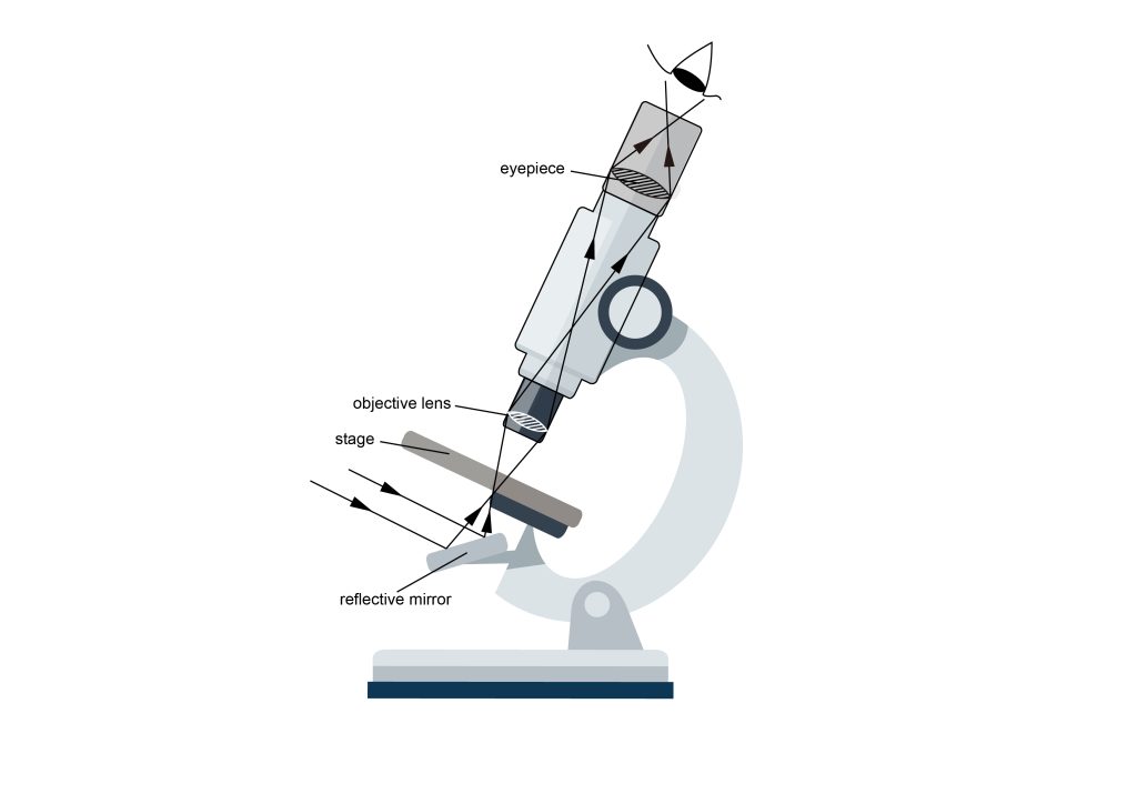
The resolution of optical microscopes is limited by the wavelength of light, typically not exceeding 0.3 micrometers. However, using ultraviolet light as the light source or immersing the specimen in oil can enhance the resolution. Optical microscopes can be classified into reflective and transmissive types based on the nature of the specimen. Reflective microscopes are used for observing opaque objects, where light is directed onto the object from above, and the reflected light enters the microscope. These microscopes are commonly employed in engineering and material science, and upright microscopes are called metallurgical microscopes. Transmissive microscopes, on the other hand, are used for observing transparent or very thin objects, allowing light to pass through them into the microscope. These microscopes are frequently utilized in biological tissue observation.
Depending on the design of their condenser and objective lenses, optical microscopes can be used to observe different types of samples. Brightfield microscopy is used to observe thin stained biological tissue samples, while darkfield microscopy, with its black background, highlights the fine details of samples, particularly useful for observing unstained samples such as live cells, employing phase contrast functionality. Additionally, techniques like differential interference contrast (DIC) microscopy are often paired with optical microscopes. Other categories include fluorescence microscopes and confocal microscopes, depending on the type of light source.
II. Five Key Features of Optical Microscopes
1. High Precision and Resolution
Optical microscopes are renowned for their high precision and resolution, capable of magnifying tiny objects to levels beyond the capability of the naked eye. This high resolution enables scientists and medical professionals to delve into the microscopic structures of biological systems, unraveling the mysteries of life. Utilizing high-quality lenses and advanced optical techniques, optical microscopes visualize the structure and morphology of cells, tissues, and minute particles, offering robust support for biological and medical research.
2. Adjustability and Flexibility
Optical microscopes feature adjustable parameters, including magnification, focal length, illumination angle, and color, allowing users to tailor observations according to specific requirements. This flexibility renders optical microscopes suitable for a variety of applications, whether in biological and medical research or industrial inspection and quality control. The ability to adjust parameters also permits researchers to explore the fine structures and properties of samples in greater depth, leading to more accurate and reliable experimental results.
3. Simple Structure and Durability
Optical microscopes typically have relatively simple structures, with fewer complex components, resulting in higher durability. Even under frequent use, optical microscopes maintain stable performance. This durability makes optical microscopes indispensable tools in laboratories, capable of enduring long-term and high-intensity work. With fewer components, maintenance and repair costs are also lower, contributing to the widespread adoption of optical microscopes in various laboratory and research settings.
4. Wide Applicability
The application of optical microscopes spans a wide range of fields, allowing observation of both fixed samples and dynamic processes of live cells and tissues. Researchers can observe essential processes such as cell growth, division, and functionality under the microscope by employing special culture dishes and staining techniques. This broad applicability positions optical microscopes as indispensable experimental tools in fields such as biology, medicine, chemistry, and physics, providing robust support for scientific research and medical diagnostics.