Ⅰ Dark field microscopy definition
Darkfield microscopy is a technique that uses oblique illumination to enhance contrast in samples that are poorly imaged under normal lighting conditions.
After direct light is blocked by an opaque diaphragm in the condenser, light passing through the specimen from an oblique angle is diffracted, refracted, and reflected into the microscope objective to form a bright image of the specimen superimposed on a dark background.
This dark background provides high contrast and can make specimens with complex backgrounds stand out with relatively little effort. Check out some examples in the image below.
Bottom: Silkworm larvae spiral and spiral tubes. When illuminated with a transmitted light microscope under darkfield conditions, the fragmented wood chips look unusually beautiful.
Top: Butterfly. Butterflies, due to the broad spectrum of wing scale patterns exhibited by members of this order, are among the most colorful members of the insect world. Digital images show the many microscopic scales that decorate much of the surface of a butterfly’s wings. Wing scales were illuminated with a darkfield sub-stage condenser and captured at low magnification (50x).
Ⅱ How darkfield microscopy works
Darkfield illumination requires blocking most of the light that normally passes through and around the specimen, allowing only oblique light to interact with the specimen.
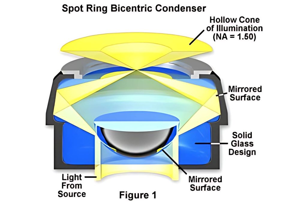
The top lens of a simple Abbe darkfield condenser is spherically concave, allowing light rays emerging from the top lens surface to form an inverted hollow cone of light focused on the specimen plane. Where there is no sample and the numerical aperture of the condenser is larger than the objective, oblique rays will cross each other and miss the objective, darkening these areas.
When a specimen (especially an unstained, non-light-absorbing specimen) is placed on a slide, oblique light rays interact with the specimen and are diffracted, reflected, and reflected by elements in the specimen, such as cell membranes, nuclei, and internal organelles. or refraction. These faint rays enter the objective lens, rendering the specimen bright against a black background.
Ⅲ Components of a darkfield microscope
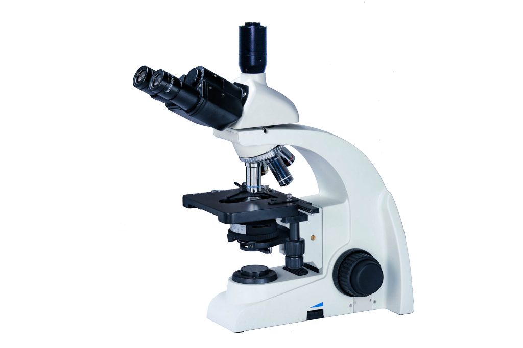
Almost any brightfield laboratory microscope can be easily converted for darkfield illumination. The simplest method is to switch off the current condenser with a condenser designed for darkfield illumination (Figure 1).
These condensers are relatively simple and provide the high numerical aperture (NA) required to create a cone of illumination for dark fields. However, switching condensers based on illumination type is impractical for daily microscopy use. Fortunately, there is a solution.
Many condensers can accommodate inserts that produce a cone of illumination. The numerical apertures of these inserts cannot be as high as dedicated darkfield condensers, so not all objectives can be used. However, inserts give you the flexibility to use multiple observation methods on the same condenser.
Ⅳ Darkfield microscopy is a great imaging technique
Generally speaking, objects imaged under appropriate darkfield lighting conditions are spectacular. Often, specimens with very low inherent contrast in brightfield microscopy shine brightly in darkfield.
Darkfield illumination is best for showing contours, edges, boundaries, and refractive index gradients. Ideal candidates for darkfield illumination include tiny living aquatic organisms, diatoms, small insects, bones, fibers, hair, unstained bacteria, yeast, cells in tissue culture, and protozoa.
Abiotic specimens include mineral and chemical crystals, colloidal particles, dust count specimens, and polymer and ceramic flakes containing small inclusions, porosity differences, or refractive index gradients.
Here are some examples of specimens captured using darkfield imaging:
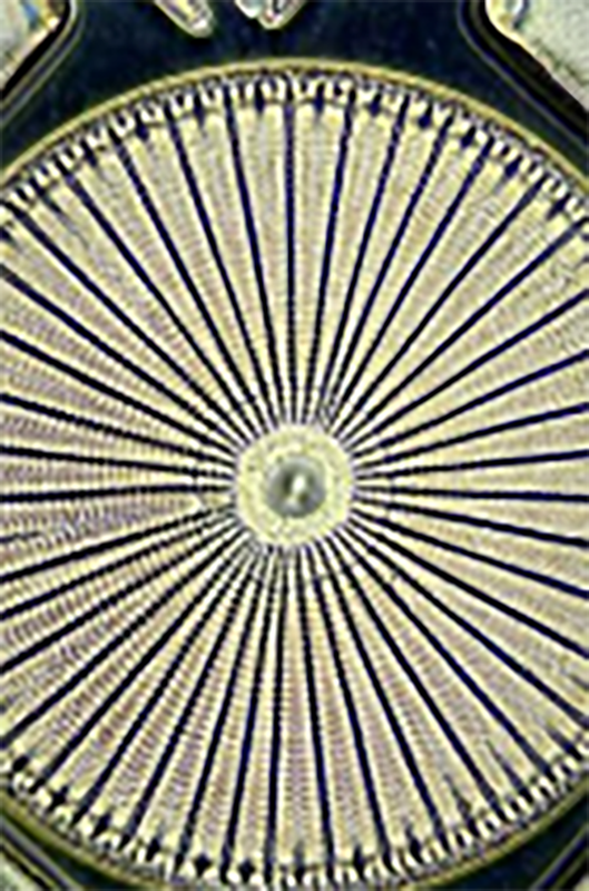
Left: This spectacular dark-field micrograph of the diatom Ehrenbergis was taken by Mortimer Abramowitz on an Olympus microscope. Illuminate the specimen with a high numerical aperture darkfield condenser, placing immersion oil between the microscope slide and the objective and condenser front lenses.
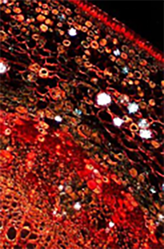
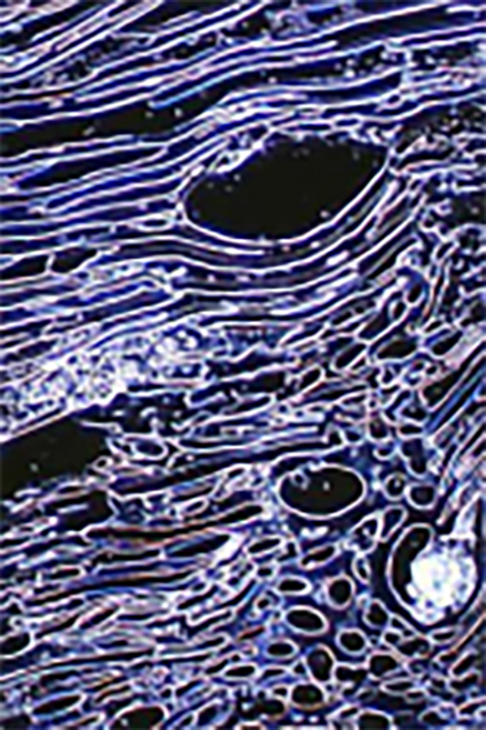
Middle: Basswood. Stained specimens are often excellent candidates for darkfield microscopy, producing beautiful images that appear in color on a dark background. This photomicrograph depicts a stained thin section of a Linden tree under darkfield illumination.
Right: Liquid crystal ribonucleic acid. This highly concentrated DNA solution undergoes a series of liquid crystal phase transitions to form a densely packed hexagonal phase. The micrograph was taken under darkfield illumination using a 10x objective of a compound optical microscope.
Unfortunately, darkfield illumination is not very useful for revealing interior details. If you want to get the most out of darkfield lighting, there are other conditions to consider.
Ⅴ What are the challenges of using darkfield microscopy?
Care must be taken when preparing specimens for darkfield microscopy because features located above and below the focal plane can scatter light and cause image degradation. The cleanliness of the slide is an important factor when imaging, but it’s even more important in darkfield because every piece of debris will be illuminated and potentially take away what you want to see.
Other challenges you may face when setting up a darkfield microscope include:
- · The lighting is insufficient to make the specimen visible, or the specimen is visible but very faintly.
Darkfield microscopy requires a lot of light to image properly because a lot of it is blocked to create a cone. So don’t hesitate to turn on the power, but be aware that some samples may not like the amount of light shining on them for an extended period of time.
- · The slide has a dark spot in the center of the field of view, but objects on the periphery are well-illuminated and look normal.
This is usually caused when the condenser is not properly aligned or focused. Centering and focusing the condenser can solve this problem. This operation is suitable for most problems where the illumination is uneven but the sample is in focus.
- · Colors appear in the background, or the background lights up unevenly with a gray cast.
This is usually caused by the sample being too thick or being mounted in contaminated media. Reloading the sample should help.
Ⅵ Advantages of using darkfield microscopy
Darkfield microscopy has many advantages. Its dark background provides high contrast, making it easy to see specimens on complex backgrounds. This technique is easy to implement because many brightfield laboratory microscopes can be configured for darkfield illumination. In addition to the benefits of microscopy, darkfield images can even look like works of art, with samples beautifully illuminated.