The materials industry is the foundation of the national economy and plays a decisive role.
The development focus of key strategic materials mainly includes special alloys, high-performance composite materials, new energy materials, new biomedical materials, electronic ceramics and intraocular lenses, rare earth functional materials, advanced semiconductor materials, etc. They are the foundation of major projects, and the era of comprehensive breakthroughs in key strategic material technologies has arrived.
In the process of material development, electron microscopes are used to characterize the structure and micromorphology of materials and analyze their impact on material properties, thereby successfully obtaining key strategic materials with excellent properties. Therefore, the electron microscope is the core equipment for materials development. So what is an electron microscope?
● What is an electron microscope?
Electron microscopy (EM) is a technique used to obtain high-resolution images of biological and non-biological specimens. It is used in biomedical research to study the detailed structure of tissues, cells, organelles and macromolecular complexes. The high resolution of EM images results from the use of electrons (which have very short wavelengths) as the source of illuminating radiation. Electron microscopy is combined with a variety of ancillary techniques (e.g., thin sectioning, immunolabeling, negative staining) to answer specific questions. EM images provide critical information about cellular function and the structural basis of cellular diseases.
There are two main types of electron microscopes – transmission electron microscope (TEM) and scanning electron microscope (SEM). Transmission electron microscopy is used to observe thin specimens (tissue sections, molecules, etc.) through which electrons can pass to create a projected image. TEM is similar in many ways to traditional (compound) optical microscopy. TEM is used to image, among other things, cell interiors (thin sections), protein molecular structure (via metallic shading contrast), viral molecular organization and cytoskeletal filaments (prepared via negative staining techniques), and the arrangement of protein molecules in cell membranes ( by freeze-fracture).
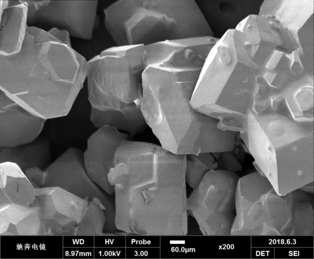
Traditional scanning electron microscopy relies on secondary electrons emitted from the surface of the sample. Due to its large focal depth, scanning electron microscopy is the EM analog of stereoscopic optical microscopy. It provides detailed images of cells and entire organism surfaces that TEM cannot provide. It can also be used for particle counting and sizing, as well as process control. It is called a scanning electron microscope because the image is formed by scanning a focused beam of electrons in a raster pattern onto the surface of the sample. Interactions of the primary electron beam with atoms near the surface cause each point in the grating to emit particles (e.g., low-energy secondary electrons, high-energy backscattered electrons, X-rays, or even photons). These can be collected with various detectors, and their relative quantities are converted into brightness at each equivalent point on the cathode ray tube. Since the size of the grating at the specimen is much smaller than the CRT’s display screen, the final image is a magnified image of the specimen. A properly equipped SEM (with secondary, backscatter, and X-ray detectors) can be used to study, for example, the morphology and atomic composition of specimens, as well as the surface distribution of immunolabels.
● Working principle of electron microscope
Theoretically, the maximum resolution that an optical microscope can achieve, d, is limited by the wavelength of photons λ that shines on the sample and the numerical aperture NA of the optical system:

Electrons have wave-particle duality, and their wave properties mean that a beam of electrons has properties similar to a beam of electromagnetic radiation. The wavelength of an electron can be found by de Broglie’s formula using the kinetic energy of the electron. Since the speed of electrons in TEM is close to the speed of light, relativistic corrections are required:

Among them, h represents Planck’s constant, m0 represents the static mass of the electron, and E is the energy of the electron after acceleration. Electrons in electron microscopes are usually emitted from a tungsten filament through the electron thermal emission process or are obtained by field electron emission. The electrons are then accelerated by the electrical potential difference and focused on the sample through electrostatic fields and electromagnetic lenses. The transmitted electron beam contains information about electron intensity, phase, and periodicity, which is used for imaging.
● Application of electron microscope
- Materials science
Electron microscopes are widely used in the field of materials science. It can be used to observe the microstructure, crystal structure, and defects of materials, analyze and characterize the physical and chemical properties of materials, help researchers understand the performance and behavior of materials, and guide the design and preparation of new materials.
▲ ① TEM image of twin crystals of face-centered cubic austenitic stainless steel ② TEM image of lattice dislocation on the atomic scale in steel ③ SEM image of porous materials
- Nanotechnology
Due to its high resolution and atomic-level resolution capabilities, electron microscopes have become an indispensable tool in nanotechnology research. It can be used to observe the morphology and structure of nanomaterials, explore the physical, chemical and biological properties at the nanoscale, and provide support for the preparation and optimization of nanodevices and nanostructures.
- Biology
Electron microscopes also have important applications in biological research. It can provide high-resolution imaging of biological samples such as cells, organelles, and biomolecules, helping scientific researchers gain an in-depth understanding of the composition and function of cells and biological structures, and study biomolecular interactions and biological processes.

●Electron microscope structure
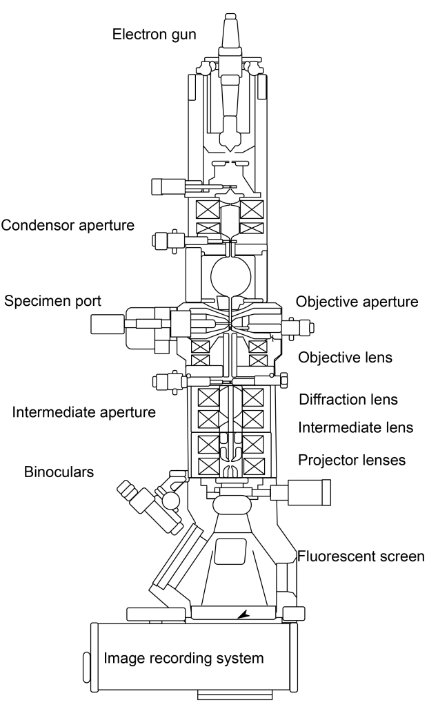
The electron microscope adopts the form of a vertically installed high vacuum column and consists of four parts: electron optical system, vacuum system and power supply system, as well as image viewing and recording system:
1. Electronic optical system
- (1) The electron optical system mainly includes components such as an electron gun, an electron lens, a sample holder, a fluorescent screen, and a camera mechanism. These components are usually assembled into a cylinder from top to bottom.
- (2) The function of the electron gun is to emit electrons as an electron source for electron microscope illumination, which is composed of a tungsten filament hot cathode, a grid, and a cathode. There are three commonly used types at present, tungsten (W) filament, lanthanum hexaboride (LaB6) filament, and field emission (Field Emission).
The electron gun can emit and form an electron beam with uniform speed, so the stability of the accelerating voltage is required to be no less than one ten-thousandth. It is the most important component of the entire electron microscope. Its performance determines the quality of the electron microscope image.
- (3) The electron lens is the most important component of the electron microscope barrel. It uses a spatial electric field or magnetic field that is symmetrical to the axis of the barrel to bend the electron trajectory toward the axis to form a focus. Its function is similar to that of a glass convex lens to focus the beam, so It’s called an electronic lens.
Most modern electron microscopes use electromagnetic lenses. The strong magnetic field generated by a very stable DC excitation current passing through a coil with pole shoes focuses the electrons.
2. Vacuum system
In order to ensure that the entire channel only interacts with the sample and does not collide with air molecules, the entire electron channel from the electron gun to the photographic plate box must be placed within the vacuum system. The general vacuum degree is 10.4 to 10.7 mm Hg.
3. Power supply system
A transmission electron microscope requires two parts of power supply: one is the high-voltage part that supplies the electron gun, and the other is the low-voltage steady-current part that supplies the electromagnetic lens.
The stability of the power supply is an extremely important indicator of the performance of the electron microscope. Therefore, the main requirement for the power supply system is to generate high and stable accelerating voltage and exciting current for each lens.
4. Image viewing and recording system
In addition to the above-mentioned power supply part, modern instruments also have automatic operation program control systems and data processing computer systems. The final image is projected on a fluorescent screen. Below the screen is a camera for recording images.
Transmission electron microscopy (TEM) Electrons can pass through the sample, so the sample must be very thin. Therefore, this electron microscope can observe the crystal structure characteristics inside the sample.
TEM contains several components, including a vacuum system for transmitting the electron beam, an electron emission source for generating the electron beam, a series of electromagnetic lenses and electrostatic disks, and equipment to move the sample into or out of the electron beam path. and movement in pathways. The imaging device then creates images using electron beams exiting the system.
The vacuum equipment used by TEM to achieve the required vacuum degree includes several stages:
- Use a rotary vane pump or diaphragm pump to evacuate the TEM to a low vacuum.
- A turbomolecular pump or diffusion pump pumps the TEM to the high vacuum required for operation.
To prevent the backing vacuum pump from running continuously and the turbomolecular pump from operating continuously, the vacuum end of the backing vacuum pump needs to be cascaded with the turbomolecular pump.
Different vacuum levels are required when using TEM (the electron gun of high-resolution TEM or field emission TEM requires a vacuum level of 10-4~10-7 Pa or even higher, while high-voltage TEM requires an extremely high vacuum level, usually It should reach 10-7~10-9Pa to prevent arc generation, especially at the cathode of TEM). If the vacuum degree of the TEM does not reach the required level, it will cause several problems. For example, the gas entering the TEM will precipitate on the sample to be observed through a process called electron beam induced precipitation, or in more serious cases. Doing so will cause cathode damage. Vacuum problems caused by the sample can be solved by using cold trap technology to absorb sublimated gases near the sample.
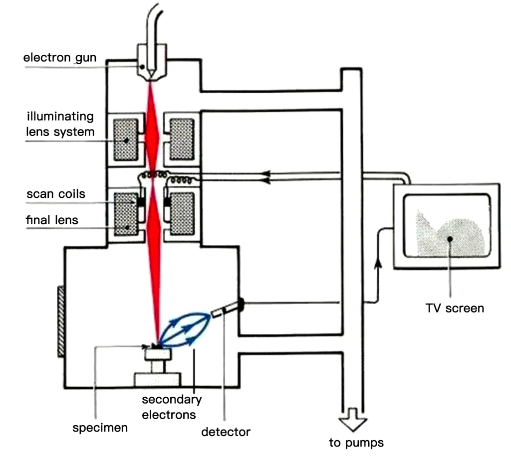
The electron beam in a scanning electron microscope (SEM) focuses on a small area of the sample as much as possible and then scans the sample line by line, so what it observes is the surface morphology and chemical composition of the surface.
The scanning electron microscope consists of three parts: a vacuum system, an electron beam system, and imaging system.
The vacuum system mainly consists of two parts: a vacuum pump and a vacuum column. A vacuum column is a sealed cylindrical container. The vacuum pump evacuates the vacuum column. There are three categories: mechanical pumps, oil diffusion pumps, and turbomolecular pumps. The combination of a mechanical pump + oil diffusion pump can meet the vacuum requirements of an SEM equipped with a tungsten gun. For SEM equipped with a field emission gun or lanthanum hexaboride gun, a combination of mechanical pump + turbomolecular pump is required.
Both the imaging system and the electron beam system are built into the vacuum column. The bottom end of the vacuum column is a sealed chamber for placing samples. The reason why vacuum is used is mainly based on the following two reasons:
- The filament in the electron beam system will quickly oxidize and fail in an ordinary atmosphere. Therefore, in addition to using a vacuum when using SEM, the entire vacuum column also needs to be filled with pure nitrogen or inert gas.
- To increase the mean free path of electrons, more electrons are used for imaging, resulting in unclear images.
If what needs to be observed is the internal structure and surface morphology of the sample, then this type of electron microscope is called Scanning Transmission Electron Microscopy (STEM). Cryo-electron microscopy is an ultra-low temperature freezing sample preparation and transmission technology (Cryo-SEM) used for scanning electron microscopy. It can directly observe liquid, semi-liquid, and electron beam-sensitive samples, such as biology, polymer materials, etc.
● The role of vacuum in electron microscopy
The vacuum environment in an electron microscope is one of the important factors to ensure the stable operation of the microscope.
- 1. Avoid gas scattering: Under vacuum conditions, gas can be excluded from the microscope, reducing the interaction between the gas and the electron beam, and avoiding the occurrence of gas scattering, thereby improving the contrast and resolution of the image.
- 2 Prevent sample oxidation: Many samples easily react with oxygen in the air under normal pressure, leading to the oxidation process. In a vacuum environment, the sample can be prevented from reacting with oxygen and maintained in its original state, thereby obtaining accurate imaging and analysis results.
- 3. Reduce collision and scattering: In a vacuum environment, the probability of collision and scattering between the electron beam and atmospheric molecules is low, which reduces the background noise in the image and improves the quality of the image.
- 4. Keep the instrument stable: The vacuum environment helps maintain the stability of the instrument, reduces interference from the external environment, and provides stable working conditions and accurate imaging results.