Electron microscope after more than 50 years of development, it has become an indispensable and important tool in modern science and technology. The application of electron microscope technology is based on the optical microscope.
Optical Microscope vs Electron Microscope: The resolution of the optical microscope is 0.2μm, and the resolution of the transmission electron microscope is 0.2nm. That is to say, the transmission electron microscope is magnified by 1000 based on the optical microscope times.
Electron microscopy is one of the most widely used microscopes today, the structure of metal atoms and even the overall arrangement of semiconductor atoms can be observed through an electron microscope. An electron microscope is currently the most widely used instrument, so what is the structure of an electron microscope?
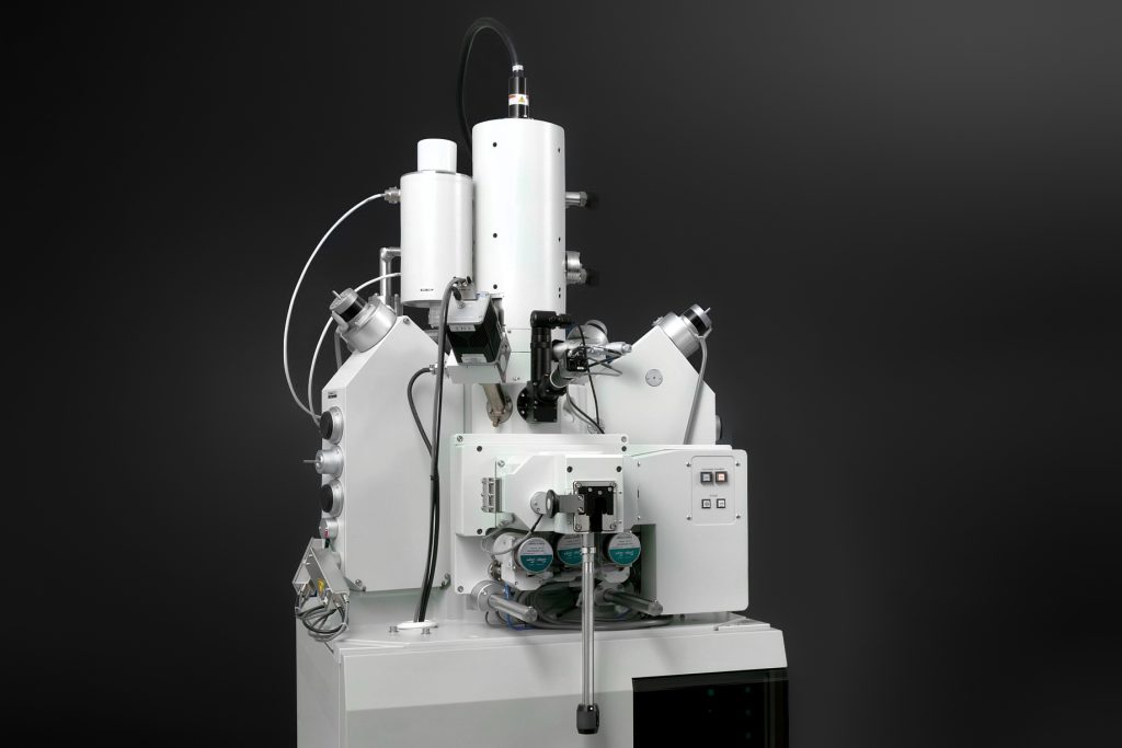
Three major components that make up an electron microscope
An electron microscope consists of a lens barrel, a vacuum device, and a power cabinet.
Components 1#: Lens Barrel
The lens barrel mainly comprises an electron source, an electron lens, a sample holder, a fluorescent screen, and a detector. It is usually assembled into a cylinder from top to bottom.
Components 2#: Electron lens
The electron lens is used to focus electrons and is the most important part of the electron microscope barrel. Typically magnetic lenses are used and sometimes electrostatic lenses are used. It uses the space electric field or magnetic field symmetrical to the axis of the lens barrel to bend the electron trajectory to the axis to form a focus, which is the same as the optical lens (convex lens) in the optical microscope to focus the beam, so it is called an electronic lens.
The focus of the optical lens is fixed, but the focus of the electronic lens can be adjusted, so the electron microscope does not have a movable lens system like an optical microscope.
Most modern electron microscopes use electromagnetic lenses to focus electrons through a strong magnetic field generated by a coil with pole shoes. The electron source consists of a cathode releasing free electrons, a grid and an anode ring accelerating electrons. The voltage difference between the cathode and anode must be high, typically between kilovolts and 3 million volts. It can emit and form an electron beam with a uniform speed, so the stability of the accelerating voltage is required to be no less than one ten-thousandth.
The sample can be stably placed in the sample holder, and there is usually a device that can be used to change the sample (such as moving, rotating, heating, cooling, elongating, etc.). Detectors are used to collect electrical or secondary signals.
Components 3#: Vacuum device
The vacuum device is used to ensure the vacuum state in the microscope so that electrons are not absorbed or deviated on its path. It consists of a mechanical vacuum pump, a diffusion pump and a vacuum valve, and is connected to the lens barrel through a suction tube.
Power supply cabinet
The power cabinet is composed of a high-voltage generator, an excitation current stabilizer, and various adjustment control units.
3 Common Types of Electron Microscopes
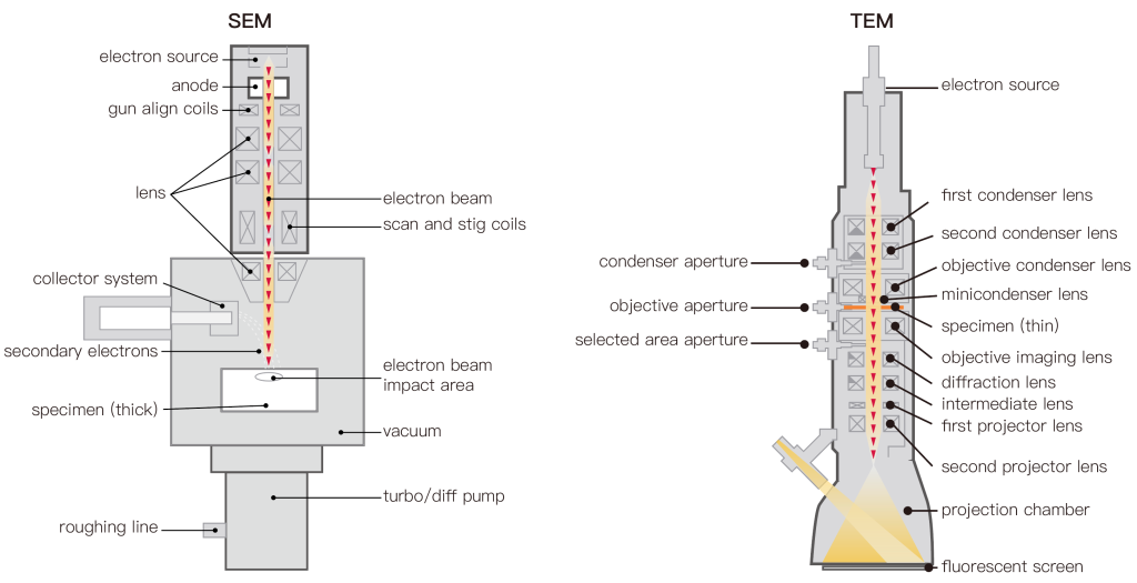
According to the structure and application, there are three common types of electron microscopes: transmission electron microscope (TEM), scanning electron microscope (SEM), and reflection electron microscope (REM).
Transmission Electron Microscope
The Transmission Electron Microscope TEM is the original type of electron microscope, and its main principle is to direct a beam of high-voltage electrons to a sample to illuminate it and produce a magnified image of the sample. Since the transmission electron microscope has the functions of in-situ observation and high-resolution imaging, it is suitable for observing the fine structures that cannot be observed by the optical microscope. For example: cell, tissue analysis, crystal structure, etc. It is often used to observe the fine material structure that cannot be distinguished by ordinary microscopes;
Scanning Electron Microscope
The scanning electron microscope is widely used, and the scanning electron microscope produces a magnified image of the specimen through a raster scanning technique. It directs a focused beam of electrons across a rectangular area of the sample, causing energy losses as the beam passes through. This energy is converted into energy such as heat, light, secondary electrons, and backscattered electrons. At this time, clear specimen image information is obtained after translation and transformation through the software system.
From the analysis of the imaging principle, the resolution of a scanning electron microscope will be slightly worse than that of the transmission electron microscope. But it has the advantage of being able to take advantage of surface treatments, create images of large samples, up to a few centimeters in size, and have a large depth of field.
Therefore, images from the SEM can better represent the real shape of the sample. It is mainly used to observe the shape of a solid surface, and it can also be combined with X-ray diffractometer or electron spectrometer to form an electronic microprobe for material composition analysis.
Reflection Electron Microscope
A reflection electron microscope involves the detection of elastically scattered electron beams reflected from a sample being examined. Reflection High Energy Electron Diffraction (RHEED) and Reflection High Energy Loss Spectroscopy (RHELS) techniques are commonly used for this type of microscopy. Generally used to study spontaneous electronic surfaces.
Transmission vs Scanning vs Reflection electron microscope
Transmission Electron Microscope (TEM), Scanning Electron Microscope (SEM), and Reflection Electron Microscope (REM) are all types of electron microscopes that use beams of electrons to visualize samples at a high resolution. However, each type of microscope has its own unique features and applications.
- Transmission Electron Microscope (TEM): In TEM, a beam of electrons is transmitted through an ultra-thin sample, producing an image that reveals the internal structure of the sample. TEM is particularly useful for studying the internal structure of biological and material samples at high magnification and resolution.
- Scanning Electron Microscope (SEM): In SEM, a beam of electrons scans the surface of a sample, producing an image that shows the surface morphology and topography of the sample. SEM is useful for studying the surface features of materials, such as the shape, size, and texture of particles, as well as the composition and structure of surfaces.
- Reflection Electron Microscope (REM): In REM, a beam of electrons is reflected off the surface of a sample, producing an image that reveals the crystal structure and surface morphology of the sample. REM is particularly useful for studying the surfaces of thin films, as well as the electronic and optical properties of surfaces.
Summary
TEM is best suited for examining the internal structure of samples, SEM is best for examining surface features, and REM is best for examining the crystal structure and surface properties of samples.