What is a Polarizing Microscope
A polarizing microscope is used to study materials with both transparent and opaque anisotropic properties, and it has significant applications in fields such as geology and other engineering disciplines.
Materials with birefringence can be clearly distinguished under a polarizing microscope. While staining methods can also be used for observation, some materials require the use of a polarizing microscope.
Reflective polarizing microscopes are essential instruments for the identification and study of materials with birefringent properties using the polarization characteristics of light. They are available for users to perform plane-polarized observation, orthogonal-polarized observation, and convergent light observation.
Basic Principles of Polarizing Microscopes
(1) Uniaxial and biaxial birefringence: When light passes through a substance, if the properties of light do not change with the direction of illumination, the substance is optically isotropic, also known as uniaxial. Examples include ordinary gases, liquids, and non-crystalline solids. If the properties of light such as speed, refractive index, absorption, polarization, and amplitude vary with the direction of illumination, the substance is optically anisotropic, also known as biaxial. Examples include crystals and fibers.
(2) Polarization of light: Light waves can be divided into natural light and polarized light based on their vibration characteristics. Natural light has vibrations in many planes perpendicular to the direction of light wave propagation, with the amplitudes of vibrations on each plane being the same. When natural light undergoes reflection, refraction, birefringence, and absorption, it can produce light waves that vibrate in only one direction, known as “polarized” or “polarized light.”
(3) Generation and effects of polarization: The most important component of a polarizing microscope is the polarization device – the polarizer and analyzer. In the past, both were made of Nicol (Nicola) prisms, natural calcite prisms. However, due to limitations in the size of crystal volumes, it was difficult to obtain large-area polarization. Therefore, artificial polarizing mirrors made of a crystal called sulphate quinoline, also known as Herapathite, are used in polarizing microscopes. These mirrors are olive green in color and, when ordinary light passes through them, produce linearly polarized light vibrating in a straight line.
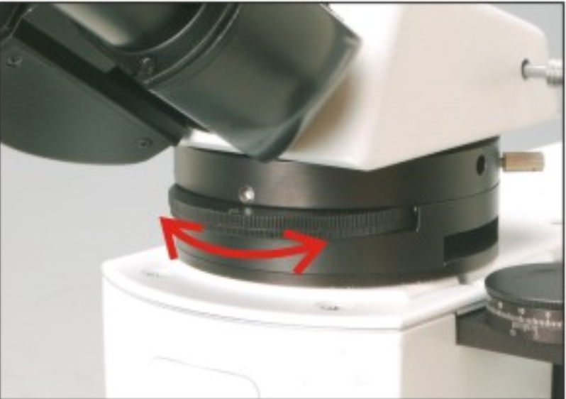
A polarizing microscope has two polarizing mirrors, one placed between the light source and the specimen called the “polarizer,” and the other placed between the objective lens and the eyepiece called the “analyzer.” The analyzer has a handle, extending mirror tube, or an intermediate attachment for easy operation, and it has a scale with rotational angles. When light passes through both polarizing mirrors, if the vibration direction of the polarizer and analyzer is parallel, i.e., in a “parallel analyzer position,” the field of view is brightest. Conversely, if they are perpendicular, i.e., in an “orthogonal analyzer position,” the field of view is completely dark. If they are inclined, the field of view shows a moderate brightness.
Therefore, in polarizing microscopy, the general principle is to place the polarizer and analyzer in an orthogonal analyzer position for observation.
(4) Birefringent substances in orthogonal analyzer position: In the orthogonal position, the field of view is dark. If the specimen is optically isotropic (uniaxial), the field of view remains dark regardless of how the stage is rotated. This is because the direction of vibration of the linearly polarized light formed by the polarizer does not change, and it remains perpendicular to the analyzer’s vibration direction. If the specimen exhibits birefringence or contains birefringent substances, the areas with birefringence become bright in the field of view. This is because when linearly polarized light emitted from the polarizer enters the birefringent substance, two linearly polarized lights with different vibration directions are generated. When these two lights pass through the analyzer, one of them is not orthogonal to the polarization direction of the analyzer, allowing it to pass through and create a bright image. The vibration directions of the two polarized lights formed when light passes through a birefringent substance depend on the type of material.
In the orthogonal position, when the stage is rotated, the image of the birefringent substance undergoes four changes in brightness during a 360° rotation, with darkening occurring every 90°. The darkening positions correspond to the positions where the two vibration directions of the birefringent substance match the two vibration directions of the polarizing mirrors, called “extinction positions.” Rotating 45° from the extinction position makes the specimen brightest, known as the “diagonal position.” This is because when the polarized light reaches the specimen at an angle of 45°, some of the light can pass through the analyzer, resulting in brightness. Based on these basic principles, polarizing microscopy can be used to determine whether a substance is isotropic (uniaxial) or anisotropic (birefringent).
(5) Interference colors: In the orthogonal analyzer position, observing birefringent substances using mixed light of various wavelengths as the light source, interference colors appear in the field of view when the stage is rotated. In addition to the brightest diagonal position, colors are also visible. The reason for the appearance of colors is mainly due to interference colors (although it is also possible that the specimen itself is not colorless and transparent). The distribution characteristics of interference colors depend on the type of birefringent substance and its thickness. This is because interference colors are caused by the dependence of the delay on the wavelength of light for different colors. If the delay in one region of the specimen is different from the delay in another region, the color of the light passing through the analyzer will also be different.
Main Uses of Polarizing Microscopes
Polarizing microscopes are important instruments for studying the optical properties of crystals and serve as the foundation for other crystal optical research methods (such as the oil immersion method, colonoscopic method, etc.).
Polarizing microscopes are essential instruments for the identification and study of materials with birefringent properties using the polarization characteristics of light. They can be used for plane-polarized observation, orthogonal-polarized observation, and convergent light observation. The method of changing ordinary light into polarized light for microscopic examination is used to distinguish whether a substance is uniaxial (isotropic) or biaxial (anisotropic).
Birefringence is a fundamental characteristic of crystals. Therefore, polarizing microscopes are widely used in fields such as minerals, chemistry, and more. In anatomy and zoology, polarizing microscopy is often used to identify structures such as bones, teeth, cholesterol, nerve fibers, tumor cells, striated muscle, and hair. The following are the application areas of polarizing microscopes.
Biological Field:
In living organisms, different fibrous protein structures exhibit obvious anisotropy, and polarizing microscopes can provide detailed information on the molecular arrangement in these fibers, such as collagen and spindle fibers during cell division.
Identification of various biological and non-biological materials: Identification of starch properties, drug component identification, fibers, liquid crystals, DNA crystals, etc.
Medical Analysis:
Detection of stones, uric acid crystals, arthritis, etc.
Geological Analysis:
In addition to common biomedical applications, polarizing microscopes can also be used for polarized detection of various minerals and crystals, widely applied in the petroleum, mining, and semiconductor industries. LED lighting and special filters can be applied in quality control and industrial analysis.
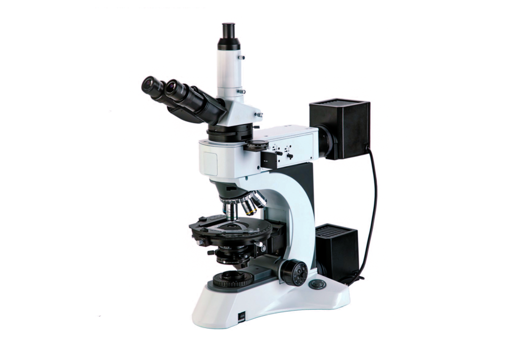
Applications of NP-800RF/TRF Series Polarizing Microscopes
Professional polarizing microscopes are used in geology, petroleum, chemical industry, polymers, liquid crystals, and other fields.
Performance of NP-800RF/TRF Series Polarizing Microscopes
- It is equipped with a polarizer that can be rotated from 0 to 90 degrees. The convergent light observation device can quickly switch between positive image mirror examination and convergent light mirror examination, with precise and fast interference image adjustment and clear images.
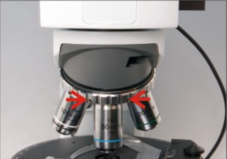
- Adjustable central five-hole objective changer.
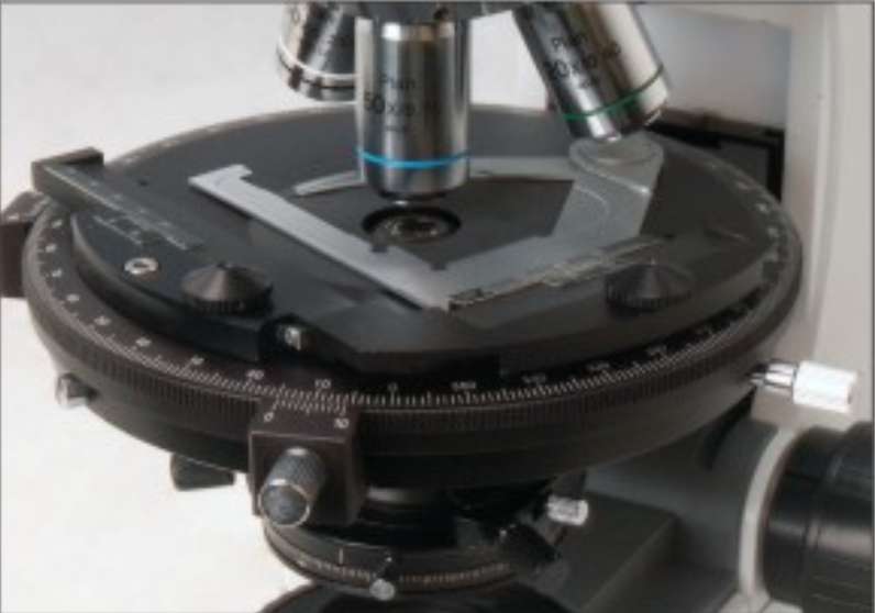
- High-precision circular rotating stage with a large surface area and convenient rotation. Equipped with a compound mechanical stage, it can control the fine movement of the sample.
Technical specifications of NP-800RF/TRF series polarizing microscope
| NP-800RF Technical Specifications | ||
| Optical System | Infinite Distance Optical System | |
| Observation System | Hinged Trinocular Tube, Inclined at 30°, Pupil Distance 48-75mm | |
| Eyepiece | EW10X/22 Flat Field Eyepiece | |
| Cross-Line Flat Field EyepieceCross Flat Field EyepieceGrid Flat Field Eyepiece | ||
| Infinity-Corrected Flat Field Achromatic Objectives | 5X/0.12/∞/- | WD 15.5mm |
| 10X/0.25/∞/- | WD 10.0mm | |
| 20X/0.4/∞/0 | WD 5.8mm | |
| 50X/0.75/∞/0 | WD 0.32mm | |
| 100X/0.8/∞/0 | WD 2mm | |
| Converter | Inward Five-Hole Converter, Center Adjustable | |
| Polarizing Device | 0°-360° Rotatable Analyzer, Minimum Scale 0.1° | |
| Conical Light Observation Device | Switchable between Positive Image Observation and Conical Light Mirror Observation, Focus Adjustable | |
| Path Length Compensator | λ Plate (First Order Red), 1/4λ Plate, Quartz Wedge Plate | |
| Rotating Round Stage | Diameter Φ170mm, Center Adjustable, 360° Scale, Scale Division 1°, Vernier Division 6′, 45° Positioned Rotation | |
| Moving Scale | Moving Range 30mm X 40mm | |
| Focusing System | Coaxial Coarse and Fine Focusing Mechanism, Focusing Range 32mm, Fine Adjustment Division 0.001mm, Coarse Movement Stroke per Rotation 37.7mm, Fine Movement Stroke per Rotation 0.1mm | |
| Polarization Device | Reflective: Fixed Type | |
| Illumination System | Reflective Illumination 24V/100W Halogen Lamp, Adjustable Brightness | |
| Filter | Φ25mm Blue Filter | |
| Φ25mm ND6 Neutral Density Filter | ||
| Φ25mm ND25 Neutral Density Filter | ||
| Accessories | Camera Accessories | |
| 0.01mm Micrometer | ||
| NP-800TRF Technical Specifications | ||
| Optical System | Infinite Distance Optical System | |
| Observation System | Hinged Trinocular Tube, Inclined at 30°, Pupil Distance 48-75mm | |
| Eyepiece | EW10X/22 Flat Field Eyepiece | |
| Cross-Line Flat Field EyepieceCross Flat Field EyepieceGrid Flat Field Eyepiece | ||
| Infinity-Corrected Flat Field Achromatic Objectives | 5X/0.12/∞/- | WD 15.5mm |
| 10X/0.25/∞/- | WD 10.0mm | |
| 20X/0.4/∞/0 | WD 5.8mm | |
| 50X/0.75/∞/0 | WD 0.32mm | |
| 100X/0.8/∞/0 | WD 2mm | |
| 40X/0.65/∞/0.17 | WD 0.54mm | |
| 100X/1.25/∞/0.17 | WD 0.13mm | |
| Converter | Inward Five-Hole Converter, Center Adjustable | |
| Polarizing Device | 0°-360° Rotatable Analyzer, Minimum Scale 0.1° | |
| Conical Light Observation Device | Switchable between Positive Image Observation and Conical Light Mirror Observation, Focus Adjustable | |
| Path Length Compensator | λ Plate (First Order Red), 1/4λ Plate, Quartz Wedge Plate | |
| Rotating Round Stage | Diameter Φ170mm, Center Adjustable, 360° Scale, Scale Division 1°, Vernier Division 6′, 45° Positioned Rotation | |
| Moving Scale | Moving Range 30mm X 40mm | |
| Condenser | NA0.9/0.25 Strain-Free Swing-Out Condenser | |
| Focusing System | Coaxial Coarse and Fine Focusing Mechanism, Focusing Range 32mm, Fine Adjustment Division 0.001mm, Coarse Movement Stroke per Rotation 37.7mm, Fine Movement Stroke per Rotation 0.1mm | |
| Polarization Device | Transmitted: 360° Rotatable, 0° Position Adjustable | |
| Reflective: Fixed Type | ||
| Reflective Illumination 24V/100W Halogen Lamp, Adjustable Brightness | ||
| Transmitted Illumination 24V/100W Halogen Lamp, Adjustable Brightness | ||
| Filter | Φ45mm Blue Filter | |
| Φ45mm ND6 Neutral Density Filter | ||
| Φ45mm ND25 Neutral Density Filter | ||
| Φ25mm Blue Filter | ||
| Φ25mm ND6 Neutral Density Filter | ||
| Φ25mm ND25 Neutral Density Filter | ||
| Accessories | Camera Accessories | |
| 0.01mm Micrometer | ||