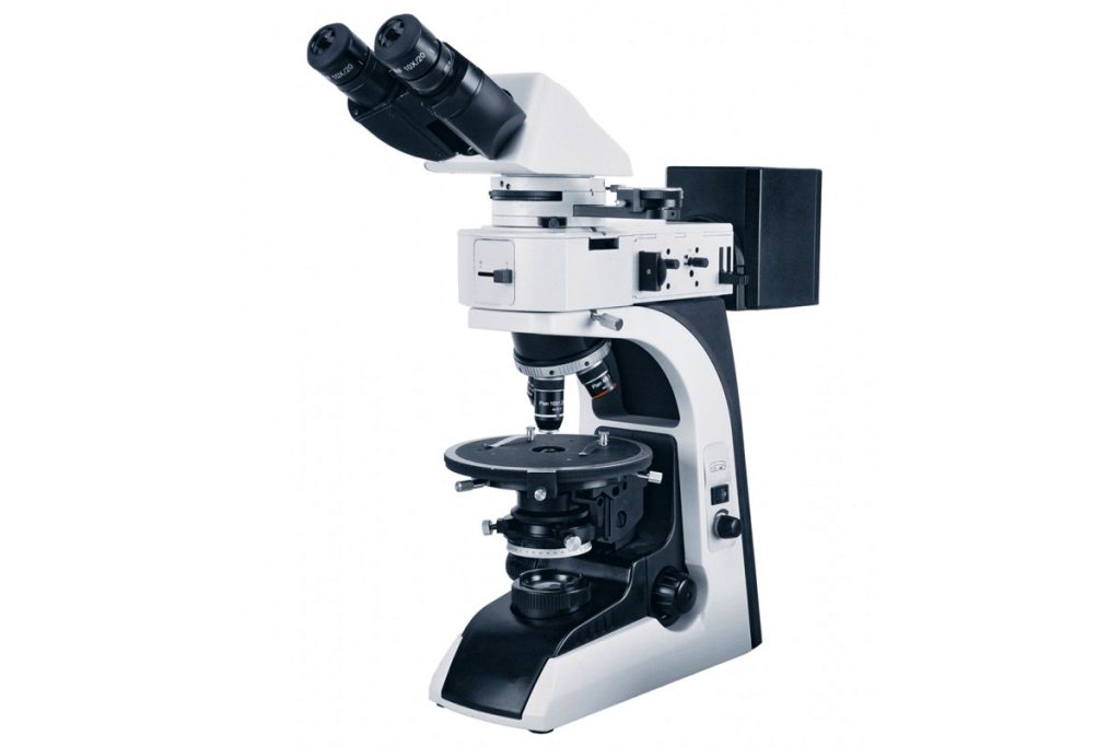
§1 What is the polarizing microscope
Polarizing microscopes detect birefringent substances, such as fibers, spindles, collagen, chromosomes, etc. This article analyzes the structure classification, technical parameters, applications, and usage precautions of polarizing microscopes.
The difference from ordinary microscopes is that there is a polarizing plate (polarizer) in front of the light source, so that the light entering the microscope is polarized light, and there is an analyzer (a polarizer with a polarization direction perpendicular to the polarizer) in the lens barrel. The stage of this kind of microscope can be rotated. When a single refractive substance is placed on the stage, no matter how the stage is rotated, because the two polarizers are vertical, no light can be seen in the microscope. When birefringent materials are introduced, the object is detected by rotating the stage because light is deflected as it passes through the material.
Any substance with birefringence can be clearly distinguished under a polarizing microscope. Of course, these substances can also be observed by staining, but for some it is impossible and a polarizing microscope must be used.
§2 Composition of polarizing microscope
There are many types of polarizing microscopes, but their structures are similar:
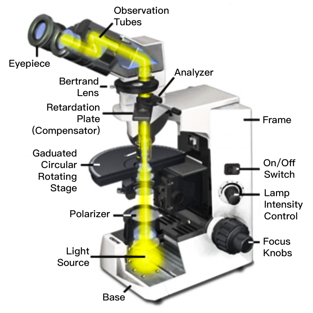
The mirror arm, bow-shaped, connects at its lower end to the mirror base and holds a lens barrel aloft.
Reflector: A round mirror, flat or concave, directs light into the microscope. For low magnification, a plane mirror suffices, while a concave mirror enhances brightness in high-magnification research.
Lower polarizer: Situated above the reflector, it polarizes natural light, denoted as PP, adjusting vibration direction via rotation.
Lock aperture: Positioned above the lower polarizer, regulates light entering the field of view with free opening and closing.
Condenser: Placed above the aperture lock, a small convex lens shapes polarized light into cones, freely adjustable in height.
Stage: A rotating circular platform with scales for precise angle reading, equipped with fixing screws and a central hole for light passage. Spring clips secure specimens.
Lens barrel: Cylinder-shaped, attaches to the mirror arm, housing an eyepiece at the upper end and an objective lens at the lower end, with additional components in between.
Objective lens: Comprised of compound lenses, including front and rear lenses, determining magnification and resolution based on numerical aperture.
Eyepiece: Consists of two lenses and accommodates crosshairs or grids, multiplying magnification alongside the objective lens.
Upper polarizer: Like the lower polarizer but perpendicular in vibration direction (AA), freely adjustable.
Bertrand lens: Positioned between the eyepiece and upper polarizer, adjustably aids viewing.
Polarizing microscopes feature additional accessories such as object stage micro-rules, mechanical stages, and tools for crystal identification. These components form optical analysis systems for crystal properties.
§3 Classification of polarizing microscopes
The main categories of polarizing microscopes are as follows:
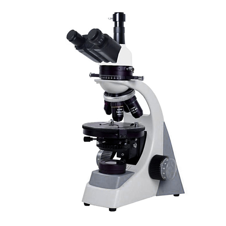
1. Binocular polarizing microscope
A binocular microscope is a microscope that allows observation with both eyes at the same time. With the development of science and technology, people found that it was inconvenient and awkward to observe with one eye, so binocular microscopes were born, which meant observing with two eyes at the same time. There were also trinocular microscopes. The third eye was usually used to connect video Imaging equipment, that is, just take a picture under the mirror and save it. Binocular observation has become a characteristic of modern visual instruments. Its function is not to feel tired after working for a long time like monocular observation. In large biological microscopes and metallographic microscopes, split-image systems are generally used to achieve binocular oblique observation.
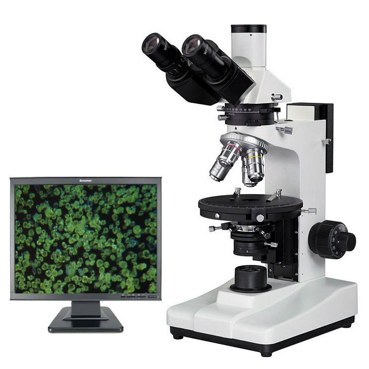
2. Digital polarizing microscope
A Digital Polarizing Microscope is a combination of a polarizing microscope host + imaging system + PC. A microscope is primarily used to study so-called transparent and opaque anisotropic materials. The physical image seen in the microscope is converted from digital to analog and imaged on the PC. The digital polarizing microscope is a high-tech product successfully developed by perfectly combining elite optical microscope technology, advanced photoelectric conversion technology, and LCD screen technology. As a result, we can reproduce the research on the microscopic field from traditional ordinary binocular observation to the monitor, thereby improving work efficiency.
3. Eight characteristics of transmission and reflection polarizing microscopes
The transmission and reflection polarizing microscope uses the principle that light will produce polarized light when reflected, refracted, birefringence or scattering under certain conditions. A polarizing filter is added to the polarizing microscope to achieve transmission and reflection observation. Features are as follows:
● eight characteristics
- a. Adopt an infinity optical system and modular functional design.
- b. Equipped with an infinity stress-free long working distance plan objective lens.
- c. Wide-angle plan eyepiece: field of view diameter Ф22mm.
- d. Coarse and micro-motion coaxial focusing mechanism, adjustable coarse and tight, with limit locking device, micro-motion grid value: 2μm.
- e. The polarizing observation device can be moved in or out of the optical path, and both the polarizer and analyzer can be rotated 360°.
- f. Rotating stage, 360° equal division scale, vernier grid value 6′, adjustable center, with locking device, effective vertical travel of the worktable up to 30mm.
- g. Wide voltage power supply (85-265V 50/60Hz). 6V30W halogen lamp lighting, adjustable brightness.
- h. The trinocular tube can freely switch between visual observation and microphotography. It can provide 100% light during photography and is suitable for low-light microscopic image shooting.
§4 Working principle of polarizing microscope
The working principle of the polarizing microscope is as follows:
1. Single refraction and birefringence:
When light passes through a certain substance, if the nature and path of the light do not change depending on the direction of illumination, the substance is optically “isotropic” and is also called a single refractor, such as ordinary gases, liquids, and amorphous substances. Solid; if light passes through another substance, the speed, refractive index, absorptivity, polarization, amplitude, etc. of the light are different depending on the direction of illumination. This substance is optically “anisotropic” and is also called a birefringent body. , such as crystals, fibers, etc.
2. The polarization phenomenon of light:
Light waves can be divided into natural light and polarized light according to the characteristics of vibration. The vibration characteristic of natural light is that there are many vibration planes on the vertical light wave transmission axis, and the amplitude distribution of vibration on each plane is the same. After natural light undergoes reflection, refraction, birefringence, and absorption, it can obtain light waves that only vibrate in one direction. This This kind of light wave is called “polarized light” or “polarized light”.
3. The generation and function of polarized light:
The polarizing microscope mainly consists of two crucial parts: the polarizer and the analyzer. Traditionally, Nicol prisms made from natural calcite were used for both, but their large size limited widespread polarization. Modern polarizing microscopes utilize artificial polarizers, typically made from quinoline sulfate crystals, known as Herapathite, exhibiting a green-olive color. These artificial polarizers efficiently produce linearly polarized light when ordinary light passes through them.
A polarizing microscope incorporates two polarizers: one positioned between the light source and the sample, known as the “polarizer,” and another placed between the objective lens and the eyepiece, called the “analyzer.” The analyzer often includes a handle for easy adjustment and a scale for angle rotation. When light passes through both polarizers, if their vibration directions align, creating a “parallel analyzer position,” the field of view appears brightest. Conversely, if the directions are perpendicular, resulting in an “orthogonal position,” the field of view is entirely dark. Intermediate brightness occurs when the directions are tilted.
This setup demonstrates that when the vibration direction of linearly polarized light aligns with the analyzer, it passes completely; when deflected, only partial passage occurs; and when perpendicular, no passage happens. Thus, during microscopy, the polarizer and analyzer ideally should be in the orthogonal analyzing position.
4. Birefringent body in orthogonal polarization position:
In the orthogonal situation, the field of view is dark. If the examined object is optically isotropic (uniaxial), regardless of how the stage is rotated, the field of view remains dark. This is because of the polarizer. This is because the vibration direction of linearly polarized light remains unchanged and is still perpendicular to the vibration direction of the analyzer.
If the examined object has birefringent properties or contains substances with birefringent properties, the field of view will become brighter in areas with birefringent properties. This is because linearly polarized light emitted from the polarizer enters the birefringent material and produces vibration directions. Two different linearly polarized lights. When these two lights pass through the analyzer, because the polarization direction of the other beam is not orthogonal to the polarization direction of the analyzer, it can pass through the analyzer, allowing the human eye to see bright colors. elephant. When light passes through the birefringent material, the vibration directions of the two polarized lights formed vary depending on the type of object.
When the birefringent material is orthogonal and the stage is rotated, the image of the birefringent material changes four times in brightness during a 360° rotation and darkens every 90°. The darkened position is the position where the two vibration directions of the birefringent material are consistent with the vibration directions of the two polarizers, called the “extinction position.” When rotated 45° from the extinction position, the examined object becomes the brightest, which is the “diagonal position,” because when the polarized light deviates from 45° reaches the object, some light will be decomposed and pass through the analyzer, so it is bright. Based on the above basic principles, polarizing microscopes can be used to determine isotropic (uniaxial) and anisotropic (birefringent) materials.
5. Interference color:
In the orthogonal analyzer position, mixed light of various wavelengths is used as the light source to observe the birefringent body. When the stage is rotated, not only the brightest diagonal position appears in the field of view, but also the color can be seen. The reason for the appearance of color is mainly caused by interference colors (of course, the object itself may not be colorless and transparent). The distribution characteristics of interference colors are determined by the type of birefringent body and its thickness, which is due to the dependence of the corresponding delay on the wavelength of light of different colors. If the delay of a certain area of the object being inspected is different from the delay of another area, then The color of the light passing through the analyzer is also different.
§5 Essential requirements for polarizing microscopes
A polarizing microscope must have the following accessories: polarizer, analyzer, compensator or phase film, special stress-free objective lens, and rotating stage.
1. Methods of polarizing microscopy
- a. Orthscope:
Also known as distortion-free microscopy, its characteristic is that it uses a low-magnification objective lens without using a Bertrand lens (Bertrand Lens), and the object under study can be directly studied with polarized light. At the same time, in order to make the illumination aperture smaller, push open the upper lens of the condenser. Normal phase microscopy is used to examine the birefringence of objects.
- b. Conoscope:
Also known as interference microscopy, it studies the interference patterns produced when polarized light interferes. This method is used to observe the uniaxial or biaxial properties of objects. In this method, a strongly converging polarized beam is used for illumination.
2. Equipment requirements for polarizing microscopes
- a.Light source:
It is best to use monochromatic light because the speed, refractive index, and interference phenomena of light vary depending on the wavelength. Normal light can be used for general microscopy.
- b.Eyepiece:
Eyepieces with crosshairs are required.
- c. Condenser:
To obtain parallel polarization, a swing-out condenser that pushes out the upper lens should be used.
- d. Bertrand lens:
An auxiliary component in the optical path of the condenser. This is an auxiliary lens that amplifies all primary phases caused by the object into secondary phases. It ensures that the eyepiece is used to observe the planar pattern formed in the back focal plane of the objective lens.
3. Requirements for polarizing microscopy
- a. The center of the stage is coaxial with the optical axis.
- b. The polarizer and analyzer should be in an orthogonal position.
- c. The film should not be too thin.
§6 Polarized microscope application
The polarizing microscope is an important instrument for studying the optical properties of crystals, and it is also the basis for other crystal optical research methods (oil immersion method, Freund’s stage method, etc.). The polarizing microscope is an essential instrument for studying and identifying birefringent substances by using the polarization characteristics of light. It can perform single polarization observation, orthogonal polarization observation, and colonoscopic observation. A method of changing ordinary light into polarized light for microscopic examination to identify whether a substance is monorefractive (isotropic) or birefringent (anisotropic).
Birefringence is a basic characteristic of crystals. Therefore, polarizing microscopes are widely used in minerals, chemistry, and other fields. In human and zoology, polarized light microscopy is often used to identify bones, teeth, cholesterol, nerve fibers, tumor cells, striated muscle and hair.
- Biological field: In living organisms, different fibrin structures display significant anisotropy, and using polarized light microscopy can provide detailed information on the molecular arrangement within these fibers. Such as collagen, spinning silk during cell division, etc.
- Identification of various biological and non-biological materials: Such as starch property identification, drug ingredient identification, fiber, liquid crystal, DNA crystal, etc.
- Medical analysis: Such as stones, uric acid crystal detection, arthritis, etc.
- Geological and mineral analysis: Study living cells of animals and plants, such as spindles in plant cells.
In addition to common biomedical applications, polarizing microscopes can also be used for polarized detection of various minerals and crystals, and are widely used in the petroleum, mining, and semiconductor industries. LED lighting and special filters can be used in quality control and industrial analysis.
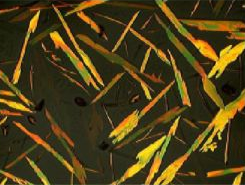
Substances under a polarizing microscope

§7 Precautions for polarizing microscope
Ensure system longevity and reliability with attention to:
- The laboratory should have three prevention conditions: shockproof (away from the source of the earthquake), moistureproof (use air conditioner, dryer), dustproof (floor covered); power supply: 220V+-10%, 50HZ temperature: 0 degrees -40 degrees.
- When adjusting the focus, be careful not to let the objective lens touch the sample to avoid scratching the objective lens.
- Do not switch the objective lens when the center of the round hole of the stage gasket is close to the center of the objective lens to avoid scratching the objective lens.
- Do not adjust the brightness from high to low, or too bright, which will affect the service life of the bulb and damage your eyesight.
- All (function) switching should be done lightly and in place.
- Adjust the brightness to the minimum when shutting down.
- Non-professionals should not adjust the lighting system (filament position lamp) to avoid affecting the imaging quality.
- When replacing the halogen lamp, pay attention to the high temperature to avoid burns; be careful not to directly touch the glass body of the halogen lamp with your hands.
- When shutting down and not in use, adjust the objective lens to the lowest state through the focusing mechanism.
- When the machine is shut down and not in use, do not cover the dust cover immediately. Wait until it cools down before covering it again. Pay attention to fire prevention.
- Optical components that are not frequently used are placed in a drying dish.
- Non-professionals should not try to clean the objective lens and other optical components. The eyepiece can be wiped with an absorbent cotton swab dipped in a 1:1 ratio (anhydrous alcohol: ether) mixed liquid and then wiped dry. Do not use other liquids to avoid damaging the eyepiece.