Introduction to Optical Microscope Observation Methods
The upright microscopes and inverted microscopes we usually use are collectively called compound microscopes, which can be used to observe smears or slices, cultured living cells, and microorganisms in water, or perform micromanipulation. The microscope host is equipped with various optical components to realize various observation methods: bright field, phase contrast, dark field, differential interference interference (DIC), etc. For different specimens, the most suitable observation method can be selected to obtain the best image.
1 bright field observation
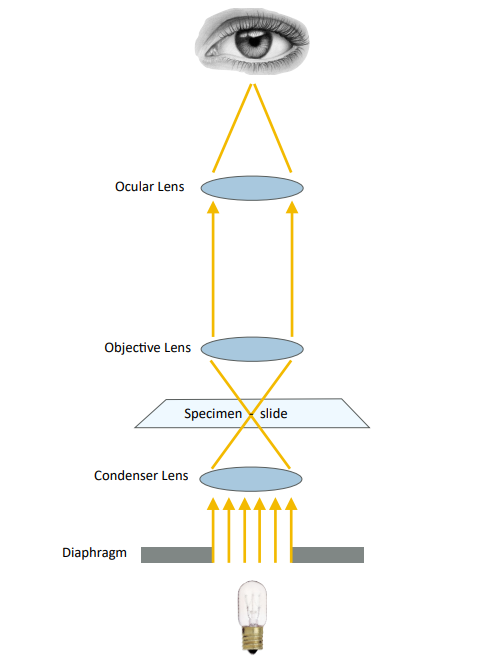
Bright field is also called bright field. Compared with dark field, bright field is directly imaged by transmitted light in the imaging process.
Commonly referred to as bright field, it means that the illumination light of the microscope light source is converged and irradiated on the specimen through the condenser lens. After being absorbed by the specimen, the remaining light enters the objective lens, and then is seen by the human eye through the eyepiece or captured by the camera through the C interface. arrive.

Blood smears, Gram-stained bacterial smears, plant slices, bone grinding slides, etc. are all observed in bright field. These types of specimens are more suitable for observation with an upright microscope. Compared with an inverted microscope, the condenser lens of an upright microscope can be very close to the specimen, and the numerical aperture is relatively large, so the overall resolution is higher than that of an inverted mirror, and the specimen can be seen more clearly.
The requirements for bright field observation are mainly clear structure and accurate color.
#advantage
- Wide range of applications and easy operation
- High brightness and uniform field of view
- Good color reproduction, flat image
#shortcoming
- Low imaging contrast (clear or unstained specimens), lack of stereoscopic effect, limited application
2 dark field observation
In the observation mode of the microscope, the dark field corresponds to the bright field, and the light is irradiated obliquely to the surface of the sample.
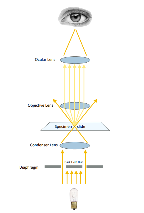
Dark field illumination is significantly different from bright field illumination in terms of optical path arrangement and lighting effect. To illustrate the effect of darkfield illumination, it is necessary to study the optical arrangement of darkfield (see figure below). The parallel light rays from the light source are blocked by the circular shading plate, and the central part of the light is blocked, and the light passing through the circular shading plate becomes a hollow cylindrical beam and enters the vertical illuminator. The vertical illuminator for dark field illumination is a circular reflector, which reflects the cylindrical light beam upwards, and casts it along the objective lens shell on the metal arc reflective surface of the reflective collector mirror, and its reflection makes the light focus on the grinding surface superior.

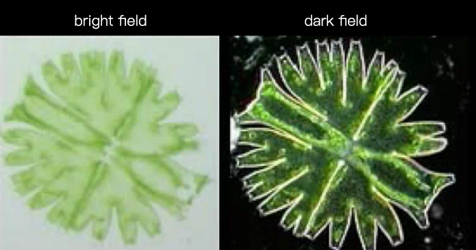
The light reflected by the reflective collector is projected on the metallographic grinding surface, because of its extremely large inclination angle, if the sample is a polished mirror, the light reflected by the sample is still reflected in the opposite direction at a large inclination angle , it is impossible to enter the objective lens. Therefore, only darkness can be seen in the eyepiece tube. Only the concave part of the sample can have light entering the objective lens, and the tissue on the sample will be reflected in the dark field of view with a bright white image, so it is called “dark field” (dark field) illumination. In most cases, the bright part of the white-on-black image obtained by dark field illumination is the black part of the black-on-white image obtained by bright field illumination.
The special accessories needed for dark field observation are dark field ring and condenser. The numerical aperture of the condenser is at least 0.2 larger than the objective lens used to achieve a better dark field effect.
Dark field observation is suitable for observing tiny aquatic organisms, fibers, hairs, bacteria, and conducting urine tests, etc.
#advantage
- Observing under dark field illumination, even very fine wear marks are easily identified.
- Can correctly identify the color of transparent non-metallic inclusions. For example, copper oxide has a bluish tint when observed in bright field under white light, but a true ruby red color can be seen when observed in dark field. Therefore, dark field observation is extremely important in the identification of non-metallic inclusions.
- Unlike bright field illumination, the light incident on the polished surface does not pass through the objective lens first, thus significantly reducing the reflection and glare caused by the light passing through the glass-air interface multiple times, and improving the contrast of the final image.
#shortcoming
- Dark field lighting must use parallel light. When using dark field observation, it is usually necessary to readjust the light source system, change the position of the collecting lens, and insert a circular shutter behind the diaphragm.
- Due to the low brightness of the object image in dark field observation, you must be very careful when photographing the light, and choose a film with a higher photosensitive speed, and the exposure time will increase accordingly.
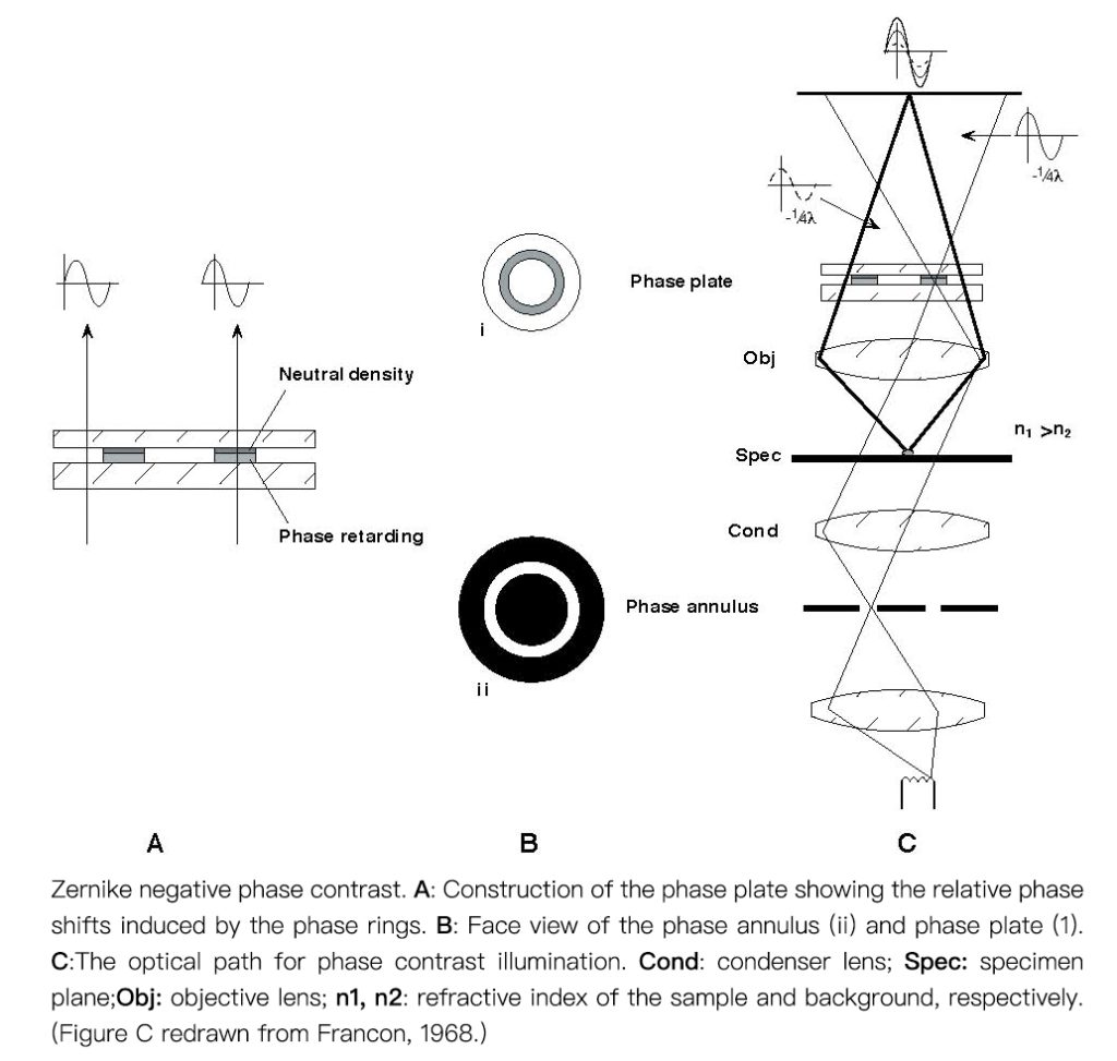
3 Phase contrast observation
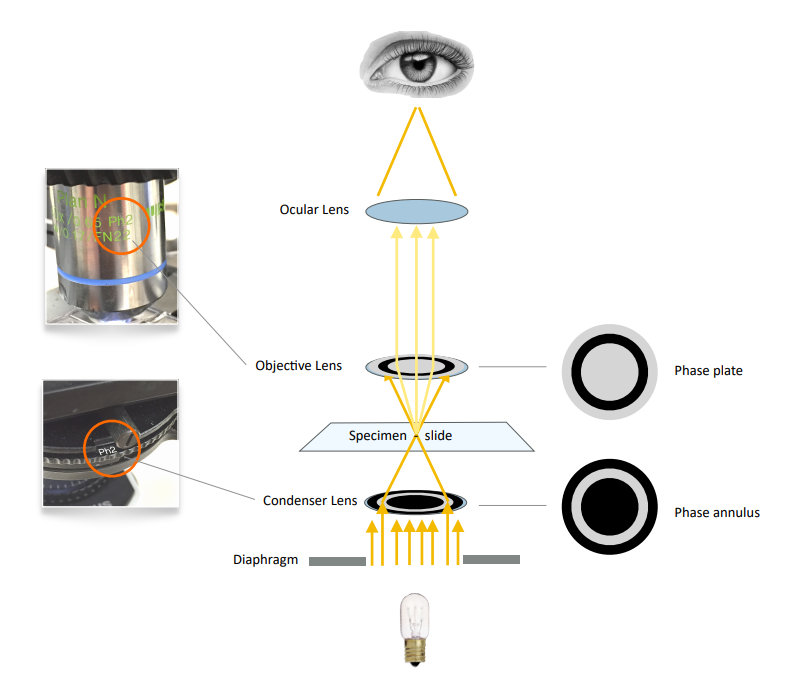
Phase contrast refers to the difference in the phase of the same light passing through media with different refractive indices. Phase refers to the position reached by the fluctuation of light at a certain time.
Generally, the phase contrast produced by the inspected object is too small to be distinguished by the naked eye, and it can only be distinguished after the phase difference is disguised as an amplitude difference (brightness and darkness difference). The phase contrast is determined by the difference of the refractive index and the thickness of the medium through which the light wave passes, which is equal to the difference of the product of the refractive index and the thickness (that is, the difference of the optical path). The phase-contrast microscope uses the difference in the optical path of the object to be inspected.
When checking the growth state of living cells in culture, the phase contrast method is commonly used for observation. Because live cells are generally not stained, and the cells are relatively transparent, it is not easy to see clearly when directly observed in bright field. The contrast method can be used to improve the contrast, and it is easier to see the state of the cells.
4 Differential interference imaging
If the cells are labeled with fluorescence, it is not suitable to observe with the phase contrast objective lens, because there is a phase ring in the phase contrast objective lens, which will block part of the fluorescence. At this time, the more suitable contrast enhancement method is differential interference imaging. Differential interference imaging can be realized by using a high-transmittance apochromatic objective lens with DIC prism and polarizer. When shooting fluorescence, the DIC component can be removed from the optical path without affecting the fluorescence imaging.
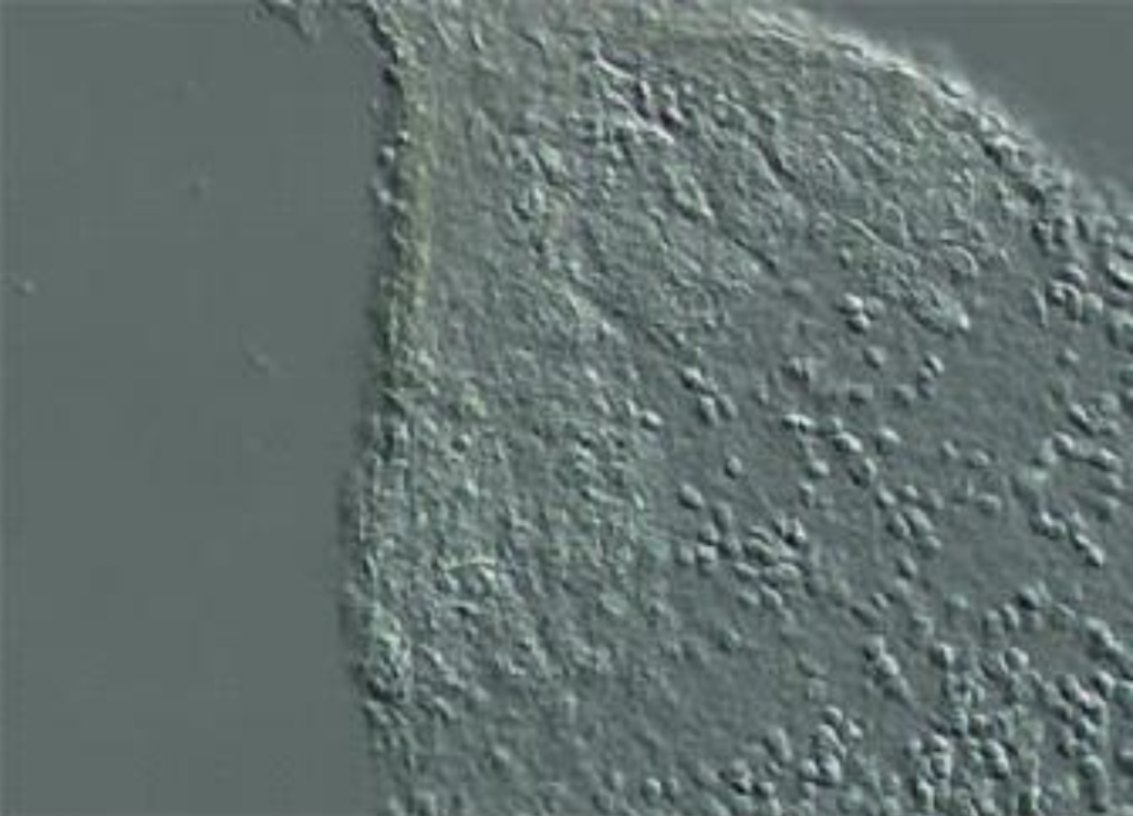
DIC presents a beautiful 3D image (not a real 3D image), no halo, and the resolution is greater than the phase difference. It is suitable for fluorescence, but it is not suitable for imaging in plastic vessels.
#advantage
- It can make the image present a three-dimensional feeling, and the observation effect is more intuitive.
- No special objective lens is needed, it works better with fluorescence observation, and can adjust the color of the background and objects to achieve the desired effect.
#shortcoming
- Not suitable for imaging in plastic vessels
Summarize
What I’m talking about today is based on the observation method of transmitted light. I hope it will be helpful to everyone.