What is Phase Contrast Microscopy?
Live, unstained cells absorb little light. Poor light absorption results in minimal differences in the intensity distribution in the image. This renders the cells little or not at all visible under brightfield microscopy. Phase contrast microscopy is an optical microscopy technique that converts the phase shift of light passing through a transparent specimen into changes in the brightness of the image. It was invented by Dutch scientist Zernike in 1935 and won the Nobel Prize in Physics in 1955.A phase contrast microscope is a special microscope especially suitable for observing objects with high transparency, such as biological slices, oil films, phase gratings, etc.
Light waves passing through these objects often only change the phase of the incident light wave without changing the amplitude of the incident light wave, because the human eye and all energy detectors can only distinguish the difference in the intensity of the light wave, that is, the difference in the amplitude, but cannot distinguish the change in phase, so it is difficult to observe these objects with ordinary microscopes.
Objects with high transparency are also called phase objects.
The phase contrast method converts the phase information of the object into the corresponding amplitude information through a spatial filter, thereby greatly improving the resolution of transparent objects. This method enables scientists to observe organic and Biological samples without the need for sample preparation, staining, or labeling.
So in this sense, the phase contrast method is an optical information processing method, and it is one of the earliest achievements of information processing, so it is of great significance in the history of optical development.
Principle of Phase Contrast Microscopy
The phase contrast microscope uses the difference in refractive index and thickness between different structural components of the object to convert the optical path difference passing through different parts of the object into the difference in amplitude (light intensity). The phase-contrast objective lens realizes the observation microscope. It is mainly used to observe living cells or unstained tissue sections, and sometimes it can also be used to observe stained samples that lack contrast.
The phase contrast microscope changes the optical path difference of the visible light passing through the specimen into an amplitude difference, thereby improving the contrast between various structures and making various structures clearly visible.
The light is refracted after passing through the specimen, deviates from the original optical path, and is delayed by 1/4λ (wavelength). If it is increased or decreased by 1/4λ, the optical path difference becomes 1/2λ, and the two beams interfere after the axis of light Strengthen, increase, or decrease the amplitude, and improve the contrast.
In terms of structure, phase contrast microscopes have four special features different from ordinary optical microscopes:
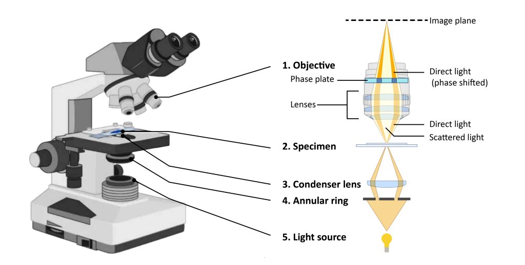
1. The annular diaphragm (annular diaphragm) is located between the light source and the condenser, and its function is to make the light passing through the condenser form a hollow light cone and focus it on the specimen.
2. Phase plate (annular phase plate) A phase plate coated with magnesium fluoride is added to the objective lens, which can delay the phase of direct light or diffracted light by 1/4λ. Divided into two types:
(1) A+ phase plate: The direct light is delayed by 1/4λ, the light waves of the two sets of light waves are added together, and the amplitude is increased. The structure of the specimen is brighter than the surrounding medium, forming a bright contrast (or negative contrast).
(2) B+ phase plate: The diffracted light is delayed by 1/4λ, and the light waves are subtracted after the two sets of light are combined on the axis, and the amplitude becomes smaller, forming a dark contrast (or positive contrast), and the structure is darker than the surrounding medium.
3. Centering telescope: The image used to adjust the annular diaphragm coincides completely with the conjugate surface of the phase plate.
How does a phase contrast microscope work?
1. Partially coherent illumination produced by a tungsten-halogen lamp is directed through the condenser and focused on a dedicated ring (labeled Condenser Ring) located in the front focal plane of the substage condenser.
2. A wavefront passing through the annulus illuminates the specimen, either through non-deviating, or diffracted and phase retarded by structure and phase gradients present in the specimen.
3. The undeviated and diffracted light collected by the objective lens is separated on the rear focal plane by the phase plate, and focused on the intermediate image plane to form the final phase contrast image observed in the eyepiece.
What are the applications of phase contrast microscopy?
Generate high-contrast images of transparent specimens such as
- live cells (usually in culture),
- Microorganisms,
- Thin tissue sectioning,
- Photolithographic patterning,
- Fibers,
- latex dispersion,
- Glass shards,
- Subcellular particles (including nuclei and other organelles).
There are many applications of phase contrast microscopy in biological research.
What are the advantages of phase contrast microscopy?
- Live cells can be observed in a natural state without pre-fixing or labeling.
- It makes highly transparent objects more visible.
- Studying objects under a phase-contrast microscope does not require special preparations such as fixation or staining, which saves a lot of time.
- Examine intracellular components of living cells with relatively high resolution.
- It enables biologists to study living cells and how they proliferate through cell division.
- Phase-contrast optics can be added to almost any brightfield microscope, provided that the dedicated phase objective fits the tube length parameters and the condenser will accept a properly sized annular phase ring. the
What are the limitations of phase contrast microscopy?
- Phase-contrast condensers and objectives add considerable cost to the microscope, so phase-contrast is not usually used in teaching laboratories except in the classroom of health professions.
- To use phase contrast, the optical paths must be aligned.
- In general, phase contrast requires more light than the corresponding brightfield observation, since the technique is based on the diminution of the brightness of most objects.