Arabidopsis thaliana, as a small model plant, has become a star in plant biology research. Its small size, short life cycle, and relatively simple genome structure make it an ideal candidate for study. In the study of Arabidopsis thaliana, stereofluorescence microscopy plays a key role. It not only helps scientists observe the internal structure of plant cells and tissues but also provides the opportunity to gain insight into the process of plant development, growth, and response to the environment. This article will discuss the application of stereofluorescence microscopy in Arabidopsis research, including its importance in gene expression analysis and signaling pathways.
01 Arabidopsis thaliana: an ideal model organism
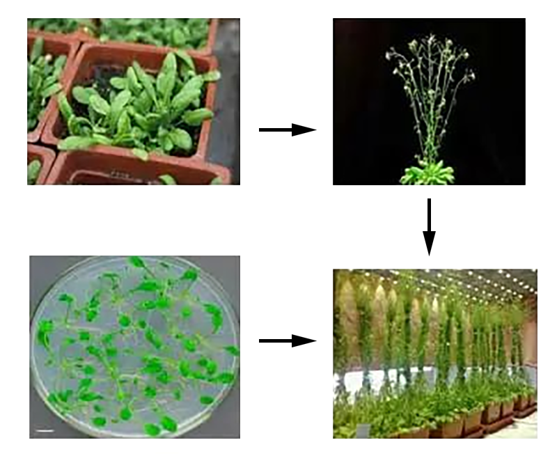
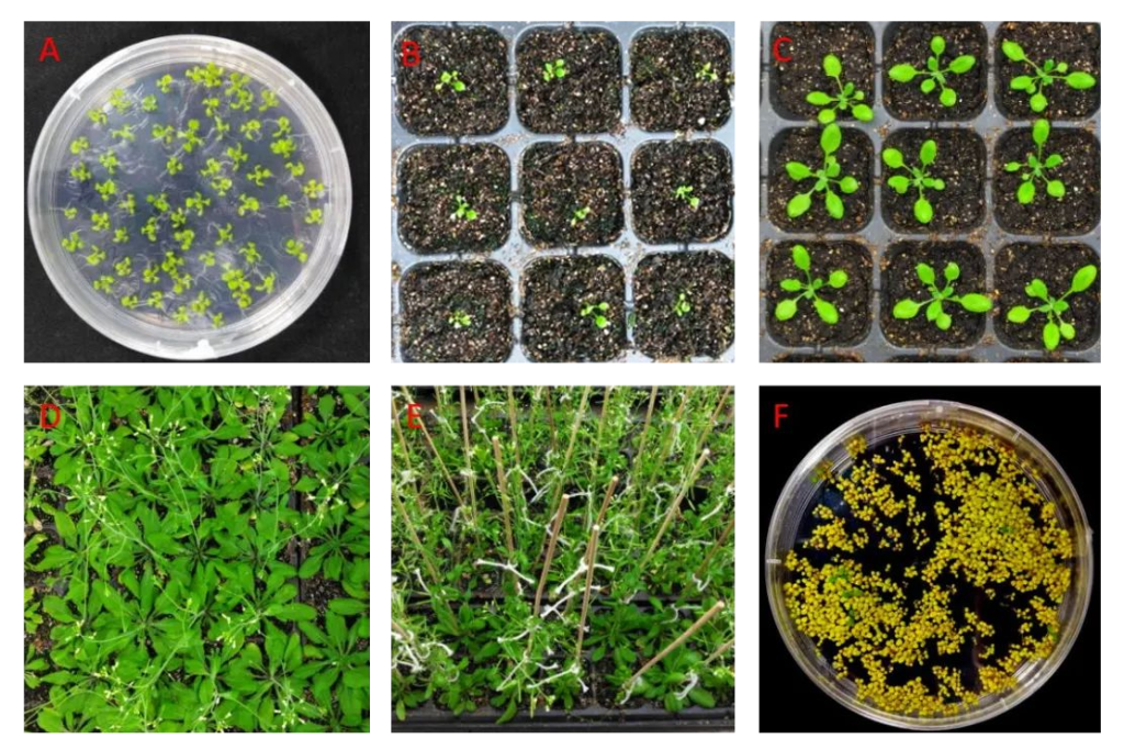
Arabidopsis is a plant in the Brassicaceae family that grows in the Middle East, especially along the Mediterranean coast. Its small size, rapid growth, and short life cycle (approximately 6 weeks) make it an ideal model plant. In addition, the Arabidopsis thaliana genome has been completely sequenced, has approximately 280 million base pairs, and contains five chromosomes. This allows researchers to easily edit and study their genes.
The adaptability of Arabidopsis thaliana also makes it an important object for ecological research. It is able to grow under different environmental conditions, thus aiding in the study of the ecological adaptability of plants. In addition, Arabidopsis plant types and flowering periods are also very diverse, providing scientists with the opportunity to study a variety of biological processes, from gene expression to growth patterns to the molecular mechanisms of resistance to stress.
02 Stereofluorescence microscopy: Exploring the internal structure of plant cells and tissues
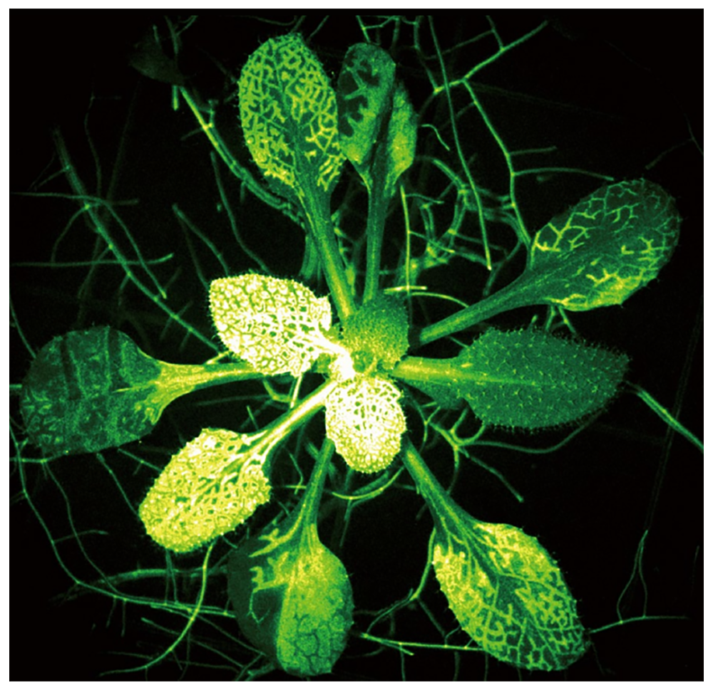
Stereofluorescence microscopy is an advanced microscopy technology that uses fluorescent signals to achieve high-resolution three-dimensional imaging, allowing researchers to delve deeply into the internal structure of plant cells and tissues. In Arabidopsis research, this technology is of critical importance because it provides the unique ability to observe cells, subcellular structures, and protein distribution, helping to shed light on plant growth and development processes.
03 Gene expression analysis
Stereofluorescence microscopy also plays a key role in gene expression analysis in Arabidopsis. Through fluorescently labeled reporter genes or protein labeling technology, scientists can visualize and quantitatively analyze the expression patterns of different genes for accurate localization of proteins in living cells and observation of dynamic changes (fluorescent protein expression, subcellular localization).
Fluorescent protein expression: Fluorescently labeled expression vectors (such as GFP or RFP) are introduced into transgenic plants to visualize the expression of target genes. Researchers can use stereomicroscopes to observe these fluorescently tagged plants to see how genes are expressed under different growing conditions.
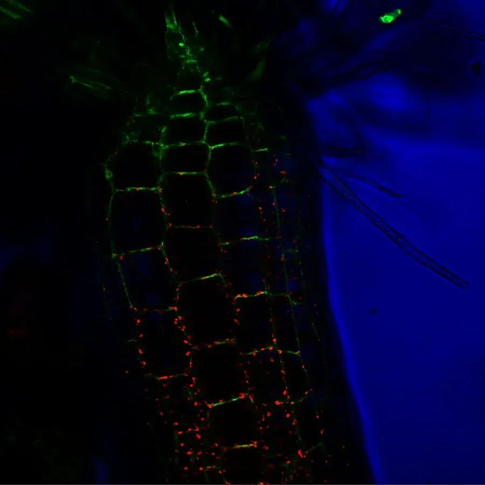
Subcellular localization: By expressing fluorescently tagged proteins in different subcellular structures, stereofluorescence microscopy can help researchers observe the intracellular localization of these proteins, such as the nucleus, chloroplasts, mitochondria, or Golgi apparatus.
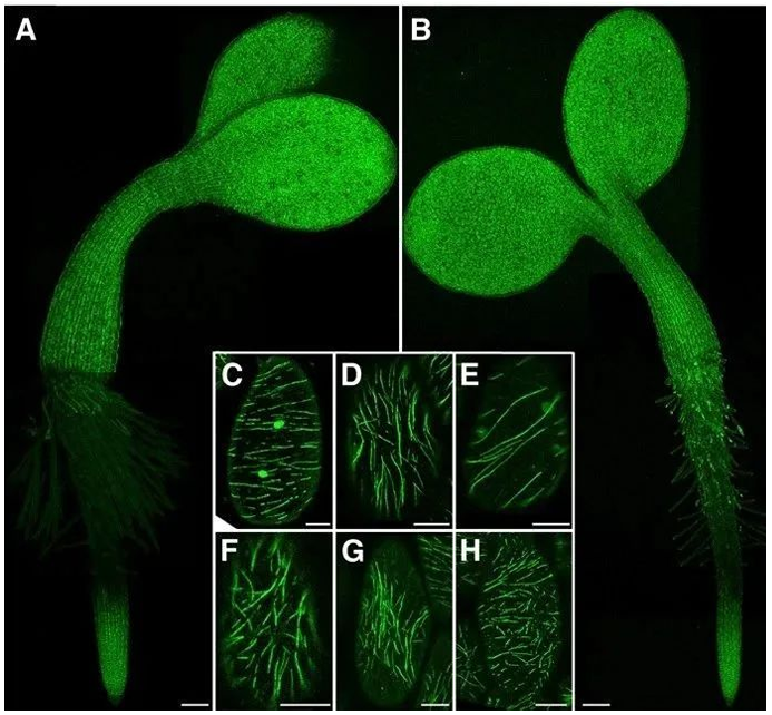
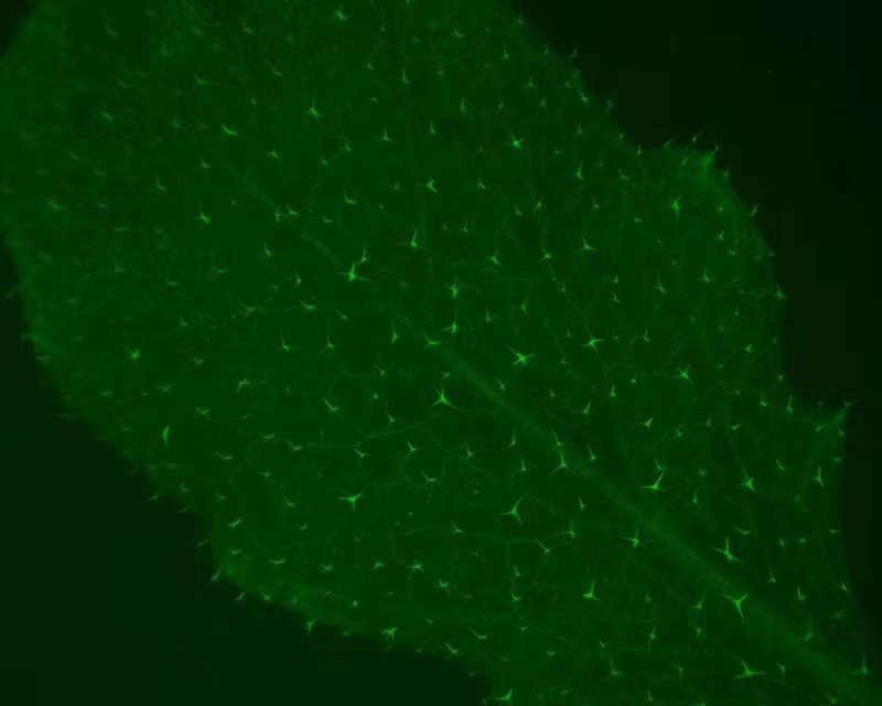
MAP65-1 is a microtubule-binding protein that plays an important role in plant cells and contributes to the stability of microtubules and the maintenance of tissue structure. By observing the distribution of microtubule skeletons in Arabidopsis leaf epidermal cells, guard cells, hypocotyl and root cells, MAP65-1 can be used to infer whether the target gene exists or whether its function is regulated.
These methods allow scientists to delve into the function of specific genes, as well as the regulation and interactions of these genes under different environmental conditions.
04 Signal transduction pathway research
Arabidopsis is also a powerful tool when studying plant signaling pathways. By introducing fluorescently labeled signaling molecules or proteins, researchers can monitor the signaling process in real-time. Some application scenarios include:
Hormone signaling: By introducing fluorescently labeled phytohormone receptors into transgenic Arabidopsis, the transmission and responses of hormone signals, such as auxin and blue light responses, can be observed.
Stress response: Researchers can use fluorescently labeled proteins to observe the physiological and molecular responses of plants under stress conditions, such as salt stress, drought, and pathogen infection.
Stereofluorescence microscopy for live imaging of plant Ca2+ signals 1
Stereofluorescence microscopy also plays a key role in the study of signaling pathways in Arabidopsis. By introducing fluorescent proteins into different protein components of signaling pathways, researchers can track protein interactions and localization during signaling. This helps to dissect the molecular mechanisms in signaling pathways and gain a deeper understanding of the process of plant growth, development and response to external stimuli.
05 Protein interaction and conformational studies
Stereofluorescence microscopy can also be used to study protein localization and conformational changes in Arabidopsis. By tagging proteins in different subcellular structures or organelles, researchers can observe the localization of these proteins to understand where they are within the cell.
Fluorescence resonance energy transfer (FRET) is a useful tool for quantifying molecular dynamics such as protein-protein interactions, protein-DNA interactions, and protein conformational changes. FRET imaging typically uses derivatives of GFP (green fluorescent protein), specifically CFP and YFP (cyan and yellow fluorescent proteins, respectively), which are attached to the protein of interest through molecular biology methods. The CFP molecules are then excited with fluorescence. Once the proteins of interest are spatially close to each other (<20 nm), CFP acts as a donor and transfers energy to YFP as an acceptor by emitting light. The researchers will observe that the blue fluorescence emitted from CFP changes to the yellow fluorescence emitted from YFP.
06 Stereofluorescence microscope
1 Introduction
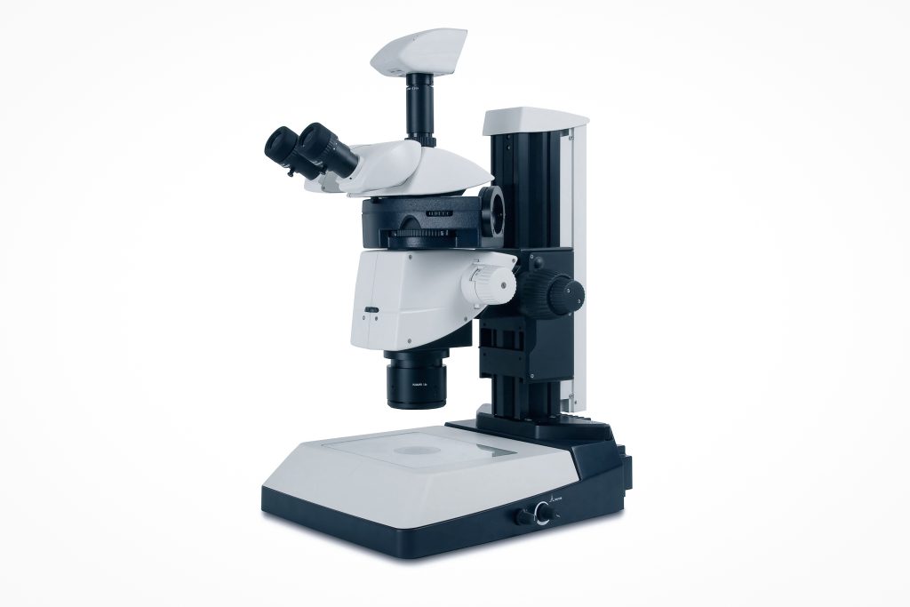
Stereofluorescence microscope is the core technology of fluorescence imaging technology in living animals. It is mainly widely used in biology, industrial detection, judicial criminal investigation and other fields. It is not only an essential tool for fluorescent anti-counterfeiting printing detection, but also an ideal instrument for mineral research.
2 Classification and microscopy
Fluorescence microscopy uses short-wavelength light to illuminate an object stained with fluorescein, causing it to be excited to produce long-wavelength fluorescence, which is then observed. Fluorescence microscopes are widely used in biology and other fields.
(1) Fluorescence microscopes are generally divided into two types: transmission and epi-illumination:
Transmission type: The excitation light comes from below the object being inspected, and the condenser is a dark field condenser, so that the excitation light does not enter the objective lens, but the fluorescence enters the objective lens. It is bright at low magnification, but becomes darker at high magnification. It is difficult to operate when immersed in oil and adjusted, especially the illumination range at low magnification is difficult to determine, but it can obtain a very dark background of the field of view. The transmission type is not suitable for non-transparent objects to be inspected.
Epi-illumination type: The transmission type has almost been eliminated. Most new fluorescence microscopes are epi-illumination type. The light source comes from above the object to be inspected and has a beam splitter in the light path, so it is suitable for both transparent and opaque objects to be inspected. Since the objective lens functions as a condenser, it is not only easy to operate, but also can achieve uniform illumination of the entire field of view from low to high magnification.
(2) Precautions for fluoroscopy:
- a. Long-term irradiation of excitation light will cause fluorescence attenuation and quenching, so shorten the observation time as much as possible. When not observing, use a baffle to cover the excitation light.
- b. When observing with an oil microscope, use “non-fluorescent oil”.
- c. Fluorescence is almost always weak and should be carried out in a darker room.
- d. It is best to install a voltage stabilizer on the power supply, otherwise, the voltage will be unstable, which will not only reduce the life of the mercury lamp but also affect the effect of the microscope.
Fluorescence microscopy is applied in many emerging biological research fields, such as gene in situ hybridization (FISH) and so on.