The eye is our powerful visual tool, but it doesn’t have the resolution to bring microscopic images into focus. This is where microscopes can help us understand everything from viruses that spark pandemics to the manufacture of increasingly miniaturized electronics, revealing how the smallest details can have a huge impact on our daily lives and future endeavors, through the microscope Observing the world can make the invisible visible, expand our perspective, change our perception, and open our minds to new possibilities.
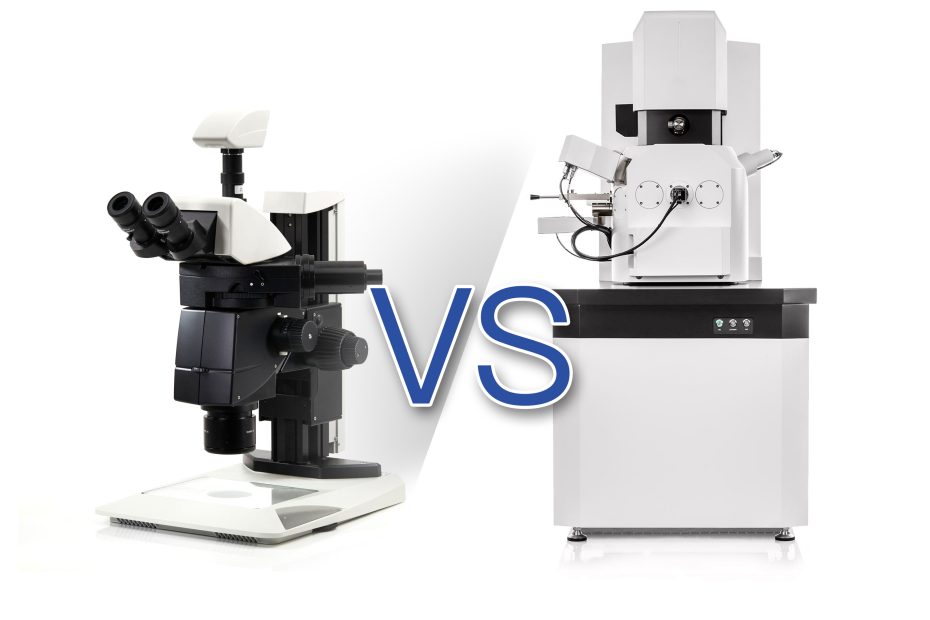
In this article, we examine two key methods: optical microscopy and electron microscopy, reveal the benefits that each technique brings, and explain their different application areas and operations.
1. Different structures and different applications
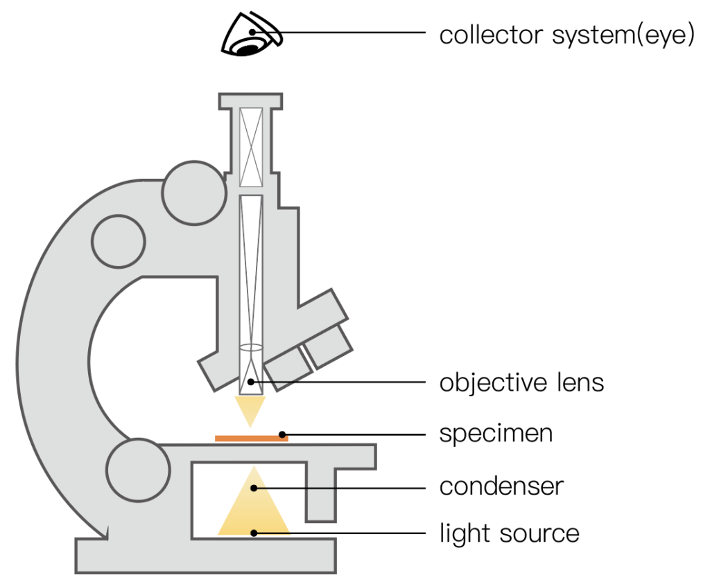
There is one major difference between optical microscopes and electron microscopes – the beam of light applied to the sample. This simple fact has major implications for the components and operation of every microscope and its applications.
- Optical Microscopy Definition: An optical microscope uses light beams with wavelengths ranging from 400nm to 650nm to allow the observer to analyze the effects of light as it hits a sample.
Ideal for general inspection purposes, an optical microscope illuminates and produces a magnified image of a specimen. The layout between optical microscopes varies by application, but typically includes a converging lens (for magnification) and a concave mirror (for auxiliary illumination). Place the sample on the stage and observe the resulting image through the eyepiece.
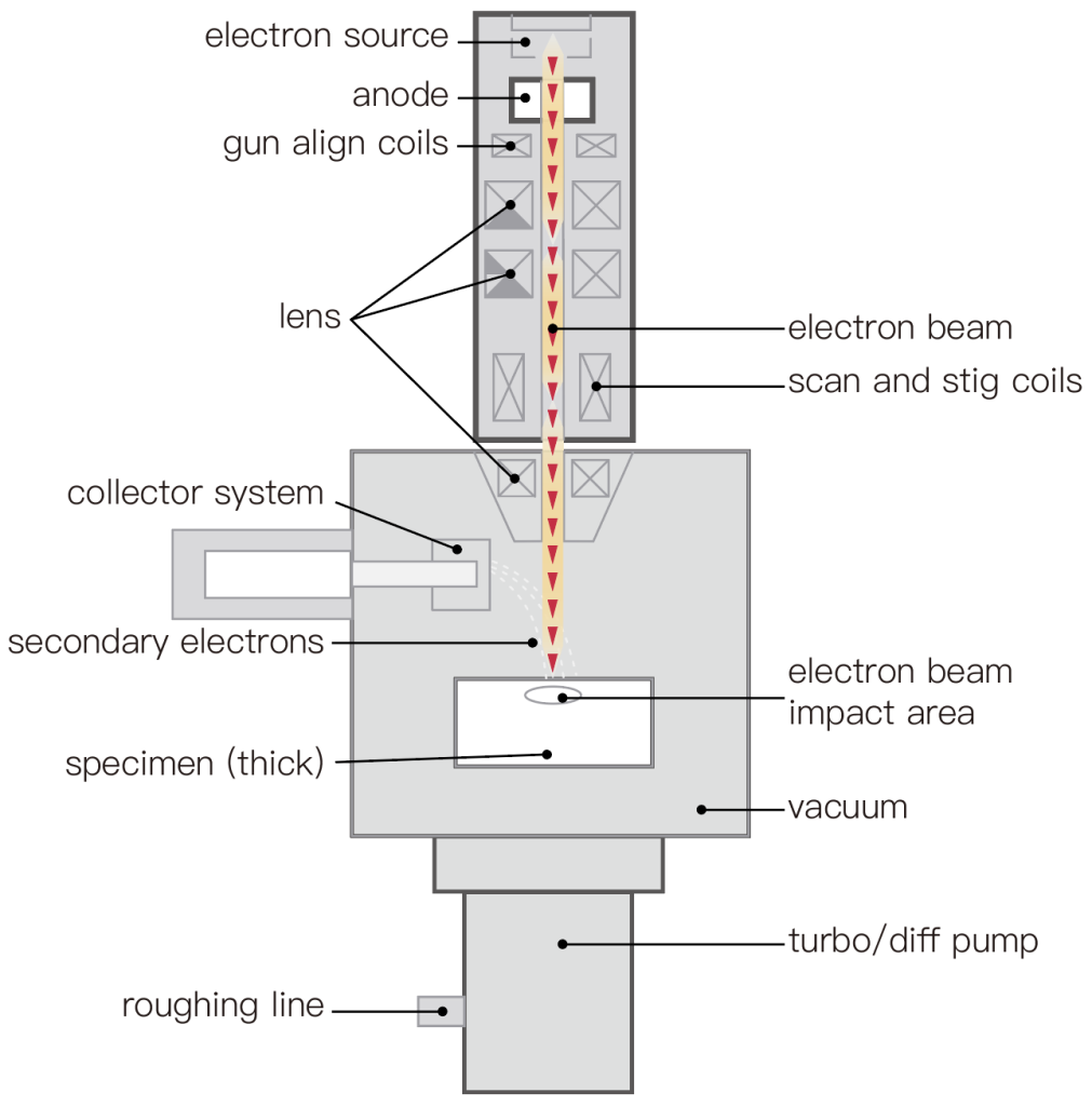
- Electron Microscope (SEM) Definition: A SEM scans a focused beam of electrons over the surface of a sample, where electromagnets are used to focus the negatively charged electrons. Interaction of the electron beam with the sample surface can affect the received image. Electrons from the sample are used to create detailed images and reveal information including texture (morphology), chemical composition, crystal structure and material orientation.
Electron microscopes typically have three types of detectors, each of which captures a different signal from the sample: secondary electron detectors (SED), backscattered electron detectors (BSED), and energy dispersive spectroscopy detectors (EDS).
Since the secondary electrons mainly interact with the sample surface and have a large reflection angle, SED can provide detailed topographical information. Backscattered electrons penetrate deep into the material and have a smaller reflection angle, so BSED provides basic morphology and basic composition information. EDS provides detailed chemical composition information.
2. The main difference between an optical microscope and an electron microscope
- Optical microscope is easy to use
Optical microscopes are easy to use, can analyze samples in air or water, and produce images with natural colors.
Electron microscopes are typically large and operate in a vacuum, which increases the time it takes to image a sample. Also, the resulting image is grayscale.
However, an electron microscope is making progress in this area, with many benchtop electron microscopes bridging the gap between light microscopy and super-resolution electron microscopy. A unique optical navigation camera displays a view of the entire sample, allowing the user to move to any location on the sample with just one click. A proprietary venting/loading mechanism supports the highest throughput, ensuring imaging times of less than 60 seconds even for large samples up to 100mm x 100mm.
- An Electron microscope has a strong resolving power
From the standard optical microscope definition, the resolving power of these systems is directly affected by the wavelength of the imaging beam, which brings clear advantages to electron microscopes. Since optical microscopes are limited to visible wavelengths, they can only provide limited magnification (approximately 1,500x) and cannot exceed a lateral resolution of approximately 200 nm and an axial resolution of 600 to 700 nm.
In contrast, electron microscopes have greater magnification and higher resolution. The most sophisticated electron microscopes can achieve approximately 100,000x magnification and subnanometer resolution, capable of imaging viruses (between 30 and 250 nm) and molecules such as proteins (10 nm) and glucose (1 nm).
- An electron microscope has a large depth of field
Due to the geometry of the imaging system, scanning electron microscopes have a much greater depth of field than optical microscopes, where the entire specimen can be brought into focus.
This is because, for an optical microscope, the depth of focus is the distance above and below the image plane where the image appears sharply. As the magnification of an optical microscope increases, the depth of focus decreases.
In contrast, an electron microscope can create a three-dimensional appearance of images of specimens. This is because of the method in which the data was acquired, in which a fine beam of electrons is scanned over the surface and the detected secondary electrons form an image with a high depth of focus.
3. Advantages and disadvantages of optical microscope and electron microscope
Advantages and disadvantages of optical microscope
Advantage:
· Low magnification (typically <1,500 x) allows for a quick, cursory inspection
· Non-destructive
· Color can be distinguished
Shortcoming:
·Limited resolution (>200 nm)
·Compared with SEM, the depth of field is smaller
·Light reflections can obscure certain features
Advantages and disadvantages of scanning electron microscope
Advantage:
· Greater depth of field compared to optical microscopes
·High magnification (typically 100,000x)
·Subnanometer resolution available
Shortcoming:
· Check times are slower
· grayscale only
4. Choose an optical microscope or an electron microscope in the application?
Compared with an optical microscope, an electron microscope shows its unique superiority. However, due to the different application technologies and fields of optical microscopes and electron microscopes, electron microscopes cannot completely replace optical microscopes.
Both light and electron microscopes have advantages and disadvantages:
- The samples of the electron microscope must be observed in a vacuum, so live samples cannot be observed;
- Electron microscopes are superior in terms of resolution and depth of focus;
- The samples of the electron microscope lack effective labels (such as the GFP of the optical microscope);
- The purchase and maintenance prices of electron microscopes are relatively high;
- Light microscopes are usually easier and faster to use.
Therefore, many people use a combination of these two imaging tools, where the light microscope is used to detect gross defects, and the electron microscope can see these defects in more detail, while viewing microscopic defects that cannot be seen with the light microscope. This two-stage approach combines the advantages associated with each detection method and provides customers with more detailed detection in less time.