Limited by the physical properties of light, the maximum resolution that an optical microscope can achieve is around 200nm (Abbe limit).
In the 1920s and 1930s, in order to see the fine structure (<100nm) inside organic cells (nuclei, mitochondria, etc.), people developed Transmission Electron Microscopy (TEM), which is based on ordinary microscopes. Instead of visible light, a focused electron beam “sees through” the specimen, greatly improving resolution.
In the 1960s, the Scanning Electron Microscope (SEM) came out, which uses a high-energy electron beam to scan a sample to the image. The electrons interact with the atoms that make up the sample and can generate signals that contain information about the sample’s surface topography, composition, and other information.
How an Electron Microscope Works
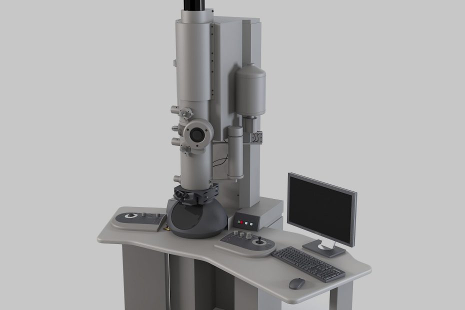
Whether it is a transmission electron microscope or a scanning electron microscope, the image is obtained by irradiating the sample with an electron beam, and its main part includes an electron source (electron gun), an electron lens, a scanning coil, a detector, etc., and these components are usually installed in a top-to-bottom Inside a high vacuum cavity/chamber (column).
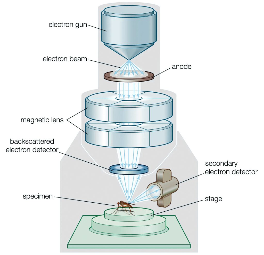
Electron gun (also known as electron source): Usually located at the top of the lens barrel, it emits electrons and forms a uniform velocity electron beam. The electron beam is focused downwards through an electron lens.
Electron lens: One of the most important components in the electron microscope barrel, it uses a space electric field or magnetic field symmetrical to the axis of the barrel to deflect the electron trajectory to the axis to form a focus, and its function is similar to that of a glass convex lens to focus the beam, so-called an electronic lens.
Most modern electron microscopes use electromagnetic lenses, which focus electrons through a strong magnetic field generated by a very stable DC excitation current passing through a coil with pole shoes.
Scan Coils: Electromagnetic coils that control and fine-tune the position of the beam.
Detector: A focused electron beam illuminates the sample on the sample stage and generates signals, which are detected by the detector and converted into an image.
The Importance of Vacuum for Electron Microscopy
Vacuum plays a very important role in electron microscopy.
Since electrons travel very slowly in the air, the vacuum system must maintain the vacuum of the electron microscope, otherwise, the molecules in the air will block the emission of the electron beam and cannot be imaged.
Two types of vacuum pumps are connected in series to obtain the vacuum in the electron microscope lens barrel. When the electron microscope is started, the first-stage rotary vacuum pump obtains a low vacuum as the pre-vacuum of the second pump; the second stage uses an oil diffusion Pump to obtain a high vacuum.
Scanning electron microscopes use high-energy electron beams to scan and image objects. In order to ensure imaging resolution, the electron beams need to be collimated and focused like beams.
And if it is not a vacuum or the vacuum degree is not enough, the high-energy electron beam hits the air molecules and is absorbed or scattered, the collimation is destroyed, and there is no way to image.
When the electron microscope is working, the entire electron beam path and the sample to be analyzed will be placed in a high vacuum environment. If the vacuum degree is not high enough, an interelectrode discharge may occur between the grid and the anode of the electron gun and the filament will be burned. Collisions of trapped air particles also cause scattering, which can prevent electrons from the electron beam from reaching the sample, or otherwise distort the analysis.
The vacuum does two things
One is to prevent air molecules from scattering electrons, resulting in the poor focus of the electron beam;
Another is to avoid the loss of electron energy after colliding with air molecules.
Vacuum Requirements for Electron Microscopes
In order to make the electrons in the electron beam move as unhindered as possible, the vacuum degree of the electron microscope usually needs to reach a high vacuum of the order of 1E-7mbar, or even an ultra-high vacuum of the order of 1E-10 mbar; Vibration levels are also critical.
Due to the small cross-section of the electron beam, the positioning accuracy on the sample is very high, and this accuracy can only be maintained in an environment with an extremely low vibration level; in addition, in order to avoid oil vapor from polluting the sample or the internal components of the microscope, it is best to use an oil-free microscope. Dry vacuum pump.
Typical Vacuum System Configuration
The image below is an example of a SEM system. From top to bottom on the left side are the lens barrel equipped with an electron gun and electron lens and the sample chamber for placing samples, and the right side of the sample chamber is a load lock.
The sample to be analyzed is placed in the inlet and outlet sample chamber, and the inlet and outlet sample chamber is pumped to a certain degree of vacuum, and then the sample is transferred to the sample chamber for analysis.
Electron microscopes with entry and exit chambers can maintain the sample chamber at a high vacuum, thereby shortening the preparation time for each inspection, increasing sample throughput, and reducing the cost of a single inspection.
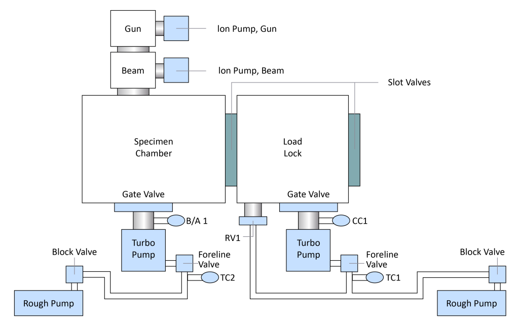
The lens barrel of the SEM is equipped with two ion pumps (Ion Pump), two sets of molecular pump (Turbo Pump) units, and multiple valves (Valve) are configured in the sample chamber and the sample chamber. Gauge to detect the vacuum degree at different positions.