All optical systems have a parameter of the depth of field.
The depth of field in photography refers to the spatial depth of the clear image obtained on the image plane, which is called the depth of field in the imaging space, or depth of field for short.
When it comes to the “depth of field” of a scanning electron microscope (SEM), it can be compared with the depth of field of photography. Both of them can obtain high-definition pictures, and they must also determine the position that needs to be focused.
When taking a photo, we bring the object of interest into focus and make it as clear as possible.
From an artistic point of view, focusing can convey the photographer’s focus to the observer, but in practice, a well-focused image can reveal a lot of detail.
But how many of the objects to be photographed are actually in focus, and how is this proportion controlled?
The part of the image that is in focus is always a plane, which means we can only image perfectly flat surfaces. Fortunately, our brains can handle that as long as parts of an object are “close enough” to the focal plane, this part is called depth of field.
1. Parameters Affecting Depth of Field
There are several parameters that affect the depth of field. Adjusting these parameters can make the picture contain more or less information.
1. The diameter of the aperture and the focusing device (camera and SEM barrel) are critical.
2. The distance between the object and the imaging device is also an extremely important factor.
In general, the farther away the object, the greater the depth of field, and more objects become clear enough for the brain to process and distinguish; the details.


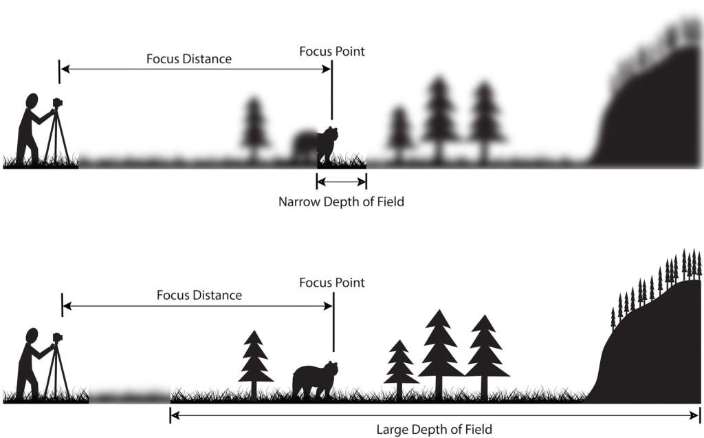
Figure 2 (a) close-up of trees – when shooting close-up, the surrounding background is blurred; (b) landscape picture – the distance is zoomed out, and the depth of field ranges from a few centimeters to several kilometers.
Both photos were taken with the same aperture and lens.
2. How the electron microscope is focused
In an electron microscope, the focal point is the position where the diameter of the cone of the incident electron beam is the smallest. The electron source emits the electron beam, and the electromagnetic lens and the end diaphragm in the column constrain the electron beam and determine the minimum beam spot size.
Resolution increases as the electron beam diameter approaches a minimum value. This value is usually achieved when a specific and optimal “working distance” – (distance between the bottom of the column and the sample) is reached.
The focal plane is the horizontal plane corresponding to the smallest diameter of the electron beam. When the electron beam is in focus, all features in the focal plane will be very sharp. Correcting focus means changing the height of the focal plane, and all features above and below the focal plane will gradually blur until they are unrecognizable.
3. How Working Distance Affects Depth of Field
Depth of field refers to the certain range of working distance within which the sharpness of the image is acceptable. The ideal working distance will give the best resolution when in focus.
However, in some cases, for example, when viewing taller samples, resolution becomes less important and depth of field has a greater influence on the results. When imaging an insect, it is critical to have all the features in the frame clearly identifiable, such as its legs and head.
The same is true for imaging electronic wiring connections, where a complete view of a sample requires the entire wire and circuit board to be in focus in the same image. In this case, a longer working distance helps to achieve a greater depth of field, resulting in images with more details clearly visible.
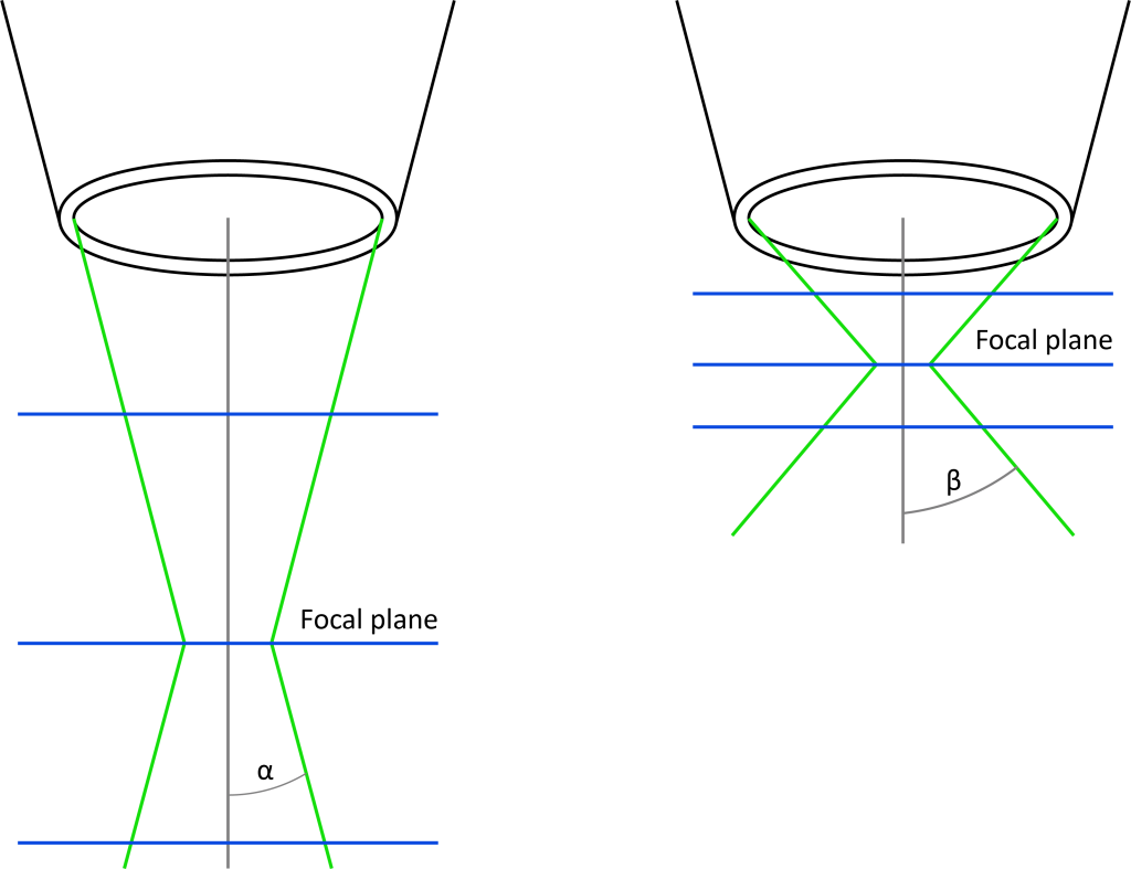
(Left image) The longer the working distance, the smaller the alpha angle, and moving away from the focal plane does not make the image too blurry.
(Right) As the working distance decreases, the beta angle becomes larger, and the diameter of the electron beam increases further away from the focal plane, so the image becomes more blurred.
Conversely, the smaller the electron beam diameter on the focal plane, the higher the image resolution.
The closer the sample is to the column, the greater the electron beam angle. This means that small deviations from the focal plane will cause the diameter of the electron beam to increase, making the image more blurred.
Conversely, the farther the sample is from the lens barrel, the smaller the electron beam angle, and the smaller the change in beam diameter caused by the deviation from the height of the focal plane, so all features at different heights can be clearly observed.
Generally, the depth of field of a scanning electron microscope can be increased from a few microns to several millimeters. By adjusting the depth of field, the features that need to be focused on can be observed in depth, and high-quality images can be obtained to achieve the best analysis results.