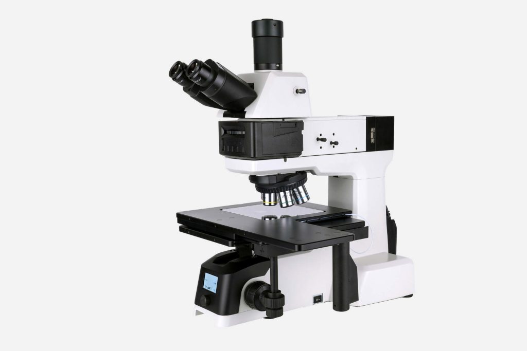
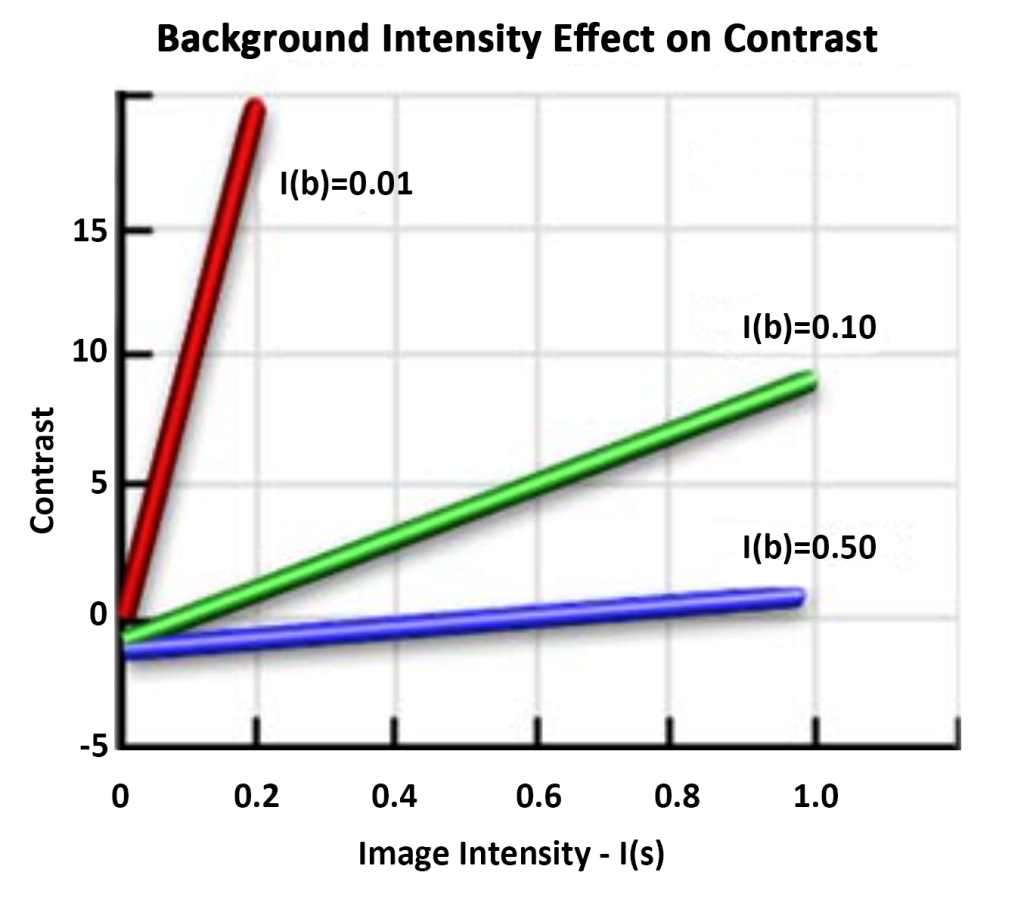
When a sample is imaged in an optical microscope, differences in intensity and/or color create image contrast, allowing individual features and details of the sample to become visible. Contrast is defined as the difference in light intensity between an image and the adjacent background relative to the overall background intensity. Generally speaking, the human eye requires a minimum contrast value of 0.02 (2%) to distinguish the difference between an image and its background. For other detectors, such as film, video cameras, or photodetectors (CCD and CMOS devices), the minimum contrast ratio is usually a different value. For each detector, the signal-to-noise ratio (noise is all the light in the optical system that has no image information) must be large enough to be able to be interpreted in terms of the formation of coherent images.
Contrast generated in a sample through light absorption, brightness, reflectance, birefringence, light scattering, diffraction, fluorescence or color changes has been a classic method for imaging samples in brightfield microscopy. The ability of a detail to stand out against the background or other adjacent details is a measure of sample contrast.
Cell staining improves contrast and allows for bright field viewing of bacteria. In addition to staining, other methods include phase contrast, differential interference contrast, darkfield, and fluorescent staining.
Tint: Enhance contrast in bright fields
Cells are stained to make them easier to see under bright-field microscopy, with each type of dye having an affinity for specific cells. Many dyes used in microbiology are positively charged and are called basic dyes, such as methylene blue, crystal violet, and safranin. Basic dyes can bind tightly to negatively charged cellular components (nucleic acids and acidic polysaccharides) and can also bind to cell surfaces, which are negatively charged. Therefore, most bacterial cells can be stained nonspecifically.
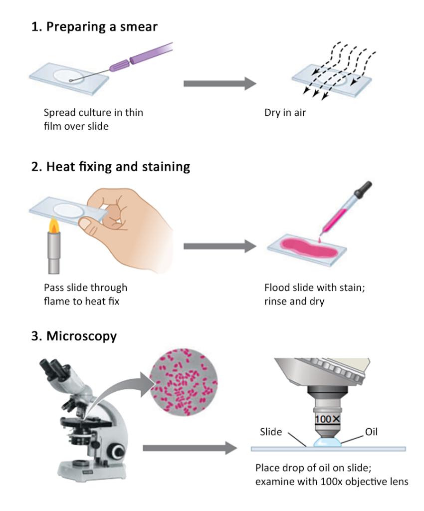
A simple staining process involves drying the cell suspension on a clean slide, soaking it in a dilute solution of basic dye for 1 to 2 minutes, rinsing it several times in water, then blotting it dry, and observing it with an oil lens.
Differential staining: Gram stain
Making different types of cells show different colors is called differential staining. An important differential staining in microbiology is Gram staining. Based on their reaction in Gram stain, bacteria can be divided into two types: Gram-negative bacteria and Gram-positive bacteria. Gram-positive bacteria are purple and Gram-negative bacteria are pink. The difference in color is caused by the different cell wall structures of the bacteria.
Phase contrast and darkfield microscopy
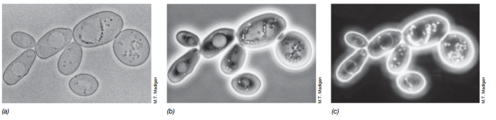
Although staining is widely used in light microscopy, it requires killing the cells. There are two types of optical microscopes that can improve the image contrast of unstained cells (that is, living cells): phase contrast microscopy and dark field microscopy. Phase contrast microscopy, in particular, is widely used in teaching and scientific research.
The basis of phase contrast microscopy: the refractive index of cells and the surrounding environment are different (that is, the material changes the speed of light). Therefore, the light passing through the cells has a different phase than the light passing through the surrounding liquid. This subtle difference is amplified by the phase contrast objective lens, and the result is in the bright background. A dark image is formed on it.
Under a dark-field microscope, light cannot penetrate the specimen. The light shines directly from the side of the specimen. Only the light scattered when it hits the specimen can reach the lens. As a result, the specimen appears bright against a dark background. Darkfield microscopy has a higher resolution than light microscopy, and some objects can be resolved with darkfield microscopy that cannot be resolved with brightfield or even phase contrast microscopy. Darkfield microscopy is also a great way to observe microbial movement.
Fluorescence microscope
Fluorescence microscopy can observe specimens that emit fluorescence. In fluorescence microscopy, cells are illuminated with one color of light from above to produce fluorescence. Only the fluorescence is visible through the filter, so the cells can glow against a black background.
Cells fluoresce, either because they contain naturally fluorescent substances, such as chlorophyll, or because they are stained with fluorescent dyes. DAPI (46-diamidino-2-phenylindole, 4′,6-diamidino-2-phenylindole) is a widely used fluorescent dye that stains cells bright blue because it interacts with Cellular DNA forms complexes. DAPI can be used to observe cells in natural habitats such as soil, water, food, or clinical specimens. DAPI staining and fluorescence microscopy are widely used in clinical diagnostics and microbial ecology to enable enumeration of bacteria in natural environments or cell suspensions.
The above-mentioned methods of improving the contrast of optical microscopes all observe microbial cells at a two-dimensional level.