Darkfield microscopy definition
Darkfield microscopy is actually a microscope that uses dark-field illumination. It is a special microscope that can distinguish the details of the sample by observing the scattered light generated when the sample is illuminated by side light. Under bright field illumination, since the illumination light source directly enters the objective lens, the scattered light from the fine structure of the sample itself cannot be distinguished. The dark field microscope can prevent the incident light from entering the observation field, making it easier to observe the scattered light of the object itself. This principle is just like in daily life, the particles of dust flying indoors are difficult to see, but in a dark room, if a beam of light slants in from the crack of the door, the dust particles will be visible.
Two Main Modes of Darkfield Microscopy
Generally speaking, compared with ordinary bright field microscopes, dark field microscopes mainly have an additional dark field ring design, so that light can only be slanted from the unobstructed parts around the dark field ring to the specimen on the slide. Since the angle of incidence of the incident light is greater than the collection angle of the collection objective, a dark background is produced. Darkfield microscopes mainly have two modes: transmission and reflection. The transmission darkfield microscope is realized by a darkfield ring, while the reflection mode is realized by a microscope lens with a darkfield ring (Figure 1 and Figure 2).
Transmission darkfield microscopy
Darkfield illumination requires blocking the central light that normally passes through and around the specimen, allowing only oblique light from all azimuths to “hit” the specimen mounted on a microscope slide. The top lens of a simple Abbe darkfield condenser is spherically concave, allowing light rays from the surface at all azimuthal angles to form an inverted hollow light cone with its apex centered in the specimen plane. If no sample is present and the numerical aperture of the condenser is larger than the numerical aperture of the objective, the oblique rays will cross and all such rays will miss entering the objective due to their inclination. The vision will appear dark.
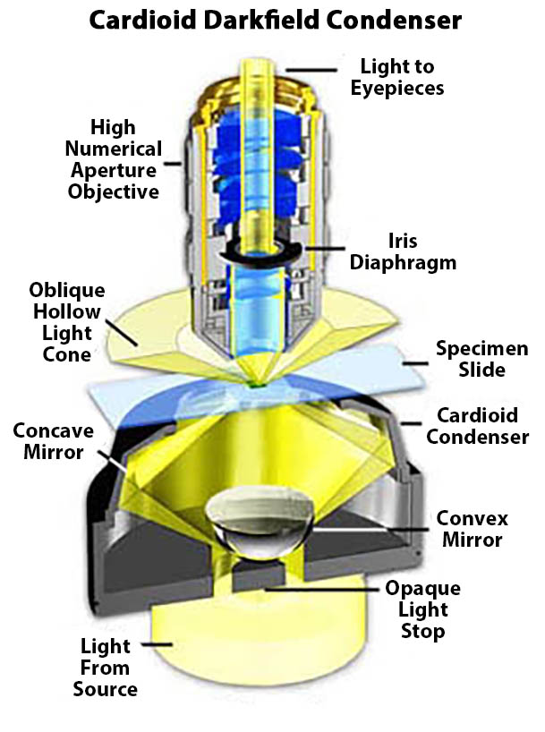
The darkfield condenser/objective pair shown in Figure 1 is a high numerical aperture arrangement that represents the most complex configuration of darkfield microscopy, which is discussed in detail below. The objective contains an internal iris diaphragm that reduces the numerical aperture of the objective to less than the numerical aperture of the inverted hollow light cone emitted by the condenser. The cardioid condenser is a reflective darkfield design that relies on internal mirrors to project an aberration-free cone of light onto the sample plane.
Reflection darkfield microscopy
Today, most darkfield reflected light microscopy objectives are infinity corrected and available in a variety of magnifications from 5x to 150x. These objectives also come in a variety of chromatic and spherical correction qualities, from plan achromatic objectives to plan fluoride BD. Most (but not all) are designed for use “dry” in the space between the objective and the sample. Some reflected light objectives are designed to focus at longer than usual working distances from the specimen. Such objectives are marked on the objective barrel as LWD (Long Working Distance), ULWD (Extra Long Working Distance), and ELWD (Extra Long Working Distance).
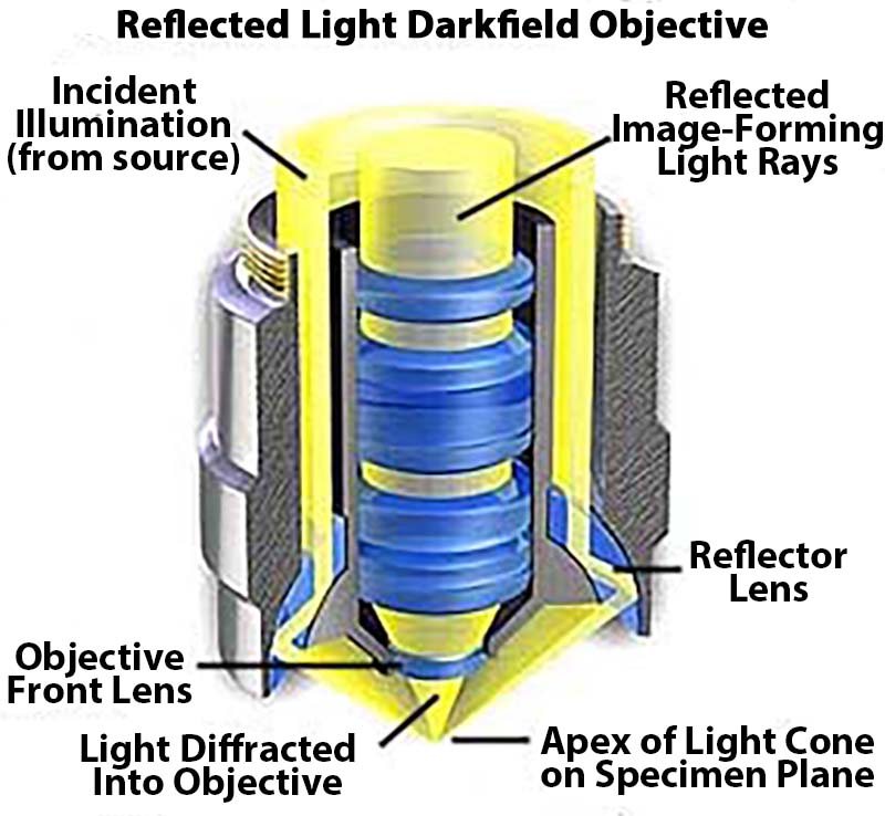
Objective designs vary between manufacturers, but the condenser section may have one of three classic designs. Reflective objectives have a single glass lens element located at the front of the objective and rely on reflections from the inner surface of the barrel to focus light onto the sample. Another important objective design is the refractive configuration, in which a series of prisms are strategically placed in a hollow outer chamber to aim and focus light onto the specimen.
The objective shown in Figure 2 is a catadioptric system that uses reflective and refractive optical elements and surfaces to create the inclined hollow cone of illumination required for viewing specimens in darkfield mode. Cylindrical light entering the hollow periphery of the objective first encounters a curved lens element that directs the light to the mirrored inner surface of the objective barrel. Light is reflected from the barrel directly through the glass element and then from the mirrored inner surface of the outer objective barrel before being refracted by a second lens element to form a hollow illumination cone. The light diffracted and refracted by the sample is then able to enter the front lens of the objective. You can explore this concept further by examining the interactive Java tutorial for Reflective Darkfield Objectives.
Applications of Darkfield Microscopy
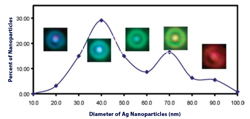
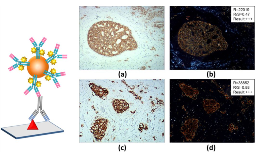
Many samples that are difficult to observe with a brightfield microscope can be observed under a darkfield microscope, such as unstained tissue specimens. Due to the localized surface plasmon resonance (LSPR) effect of metal nanoparticles, the scattered light can be directly observed through dark field microscopy. The LSRP of metal nanoparticles is affected by multiple factors such as morphology, size and surrounding environment, and displays different colors under a dark field microscope. Silver nanoparticles of different sizes from 20 nm-90 nm will display scattered light consisting of blue-violet, blue, green, and red (Figure 2). Since the scattering cross section of metal nanoparticles is very large, the light scattered by a metal nanoparticle of about 80 nm is equivalent to the light intensity produced by 5 million single fluorescent molecules, which is 1000 times stronger than a 100 nm fluorescent microsphere and stronger than a Quantum dots are 105 times stronger [2]. The interaction of individual metal nanoparticles with cells, as well as the mechanism by which nanoparticles enter cells, can therefore be tracked using dark-field microscopy. Dark-field scattering of gold nanoparticles has also been innovatively used in combination with enzyme catalysis to realize immunohistochemical detection of pathological tissues, which is expected to develop new technologies for quantitative pathological detection.