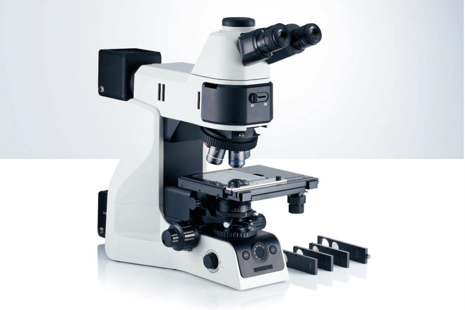
Optical microscopes use visible light and a series of lenses to magnify specimens and make detailed observations. Bright-field microscopy uses light to create dark images against a bright background. Dark-field microscopy works the opposite of bright-field microscopy, the entire field of view appears dark when no specimen is present, and once a specimen is placed, it appears bright against this dark background.
Bright-field microscope
Bright-field microscopy is a routine technique. Suitable for observing the natural color of specimens or observing stained samples. Specimens appear darker against a bright background. To increase contrast, the condenser should be turned off. This will reduce resolution but has a positive tradeoff. Diffraction increases and the edges of the sample become darker (even though they are not actually). These are artifacts, but it is difficult to see specimens (such as bacteria) more clearly.
Suitability for Amateur Microscopy: High. This is a standard technique available in all microscopes.
Dark-field microscope
Dark-field microscopy shows specimens bright against a dark background. A bright-field microscope with a condenser with a filter holder can be easily converted to a dark-field by placing a diaphragm-cut filter into the filter holder. Filters block direct light from the microscope. Specimens appear bright because they reflect the microscope’s light into the objective lens. One of the disadvantages of dark-field is that it is very sensitive to dust. A small amount of dust is already lit on a dark background.
Suitability for Amateur Microscopy: High. Converting a bright-field microscope to a dark-field is easy if your microscope has a condenser with a filter holder. You need to insert a (homemade) dark field patch stop filter.
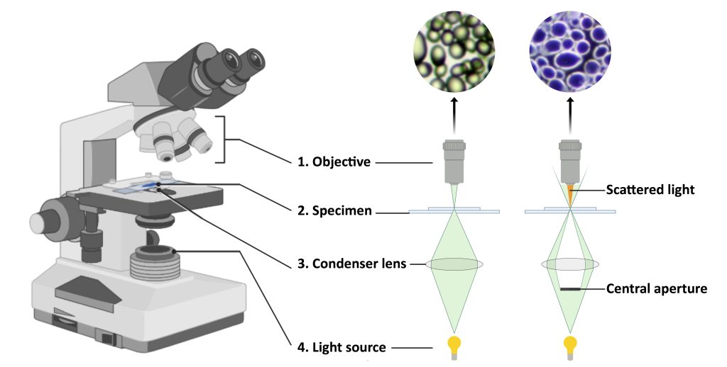
Both brightfield and dark-field microscopes are light microscopes that use light to view and magnify a specimen, but the similarities end there. The working principles of the two microscopes are completely different.
Bright-field Microscopy vs. Dark-field Microscopy
| Range | Bright-field | Dark-field |
| Image formation | works on the principle of absorption of light. The light is focused on the specimen, it absorbs the light, viewing a dark contrast image against a bright background | When there is no specimen, the entire field of view appears dark, once a specimen is placed, it appears bright against this dark background |
| Picture type | Appears hazy or dark (depending on the stain used) | Bright image on a dark background |
| Specimen Type | View fixed specimens and usually stain them | View all live specimens even if unstained |
| Specimen preparation | Complicated as specimens need to be stained | No stain required |
| Specimen color | Appears hazy or dark (depending on stain used) | Specimen appears bright |
| Specimen background | Very bright background | Very dark background |
| Type of condenser used | Abbe condenser, variable focal length condenser, achromatic condenser | Abbe condenser, parabolic condenser, cardioid condenser. |
| Viewing range | It shows the internal structure and morphological structure of the specimen | It shows the external structure of the specimen |
| Cost | They are cheap | They are more expensive |
| Opaque Disc | Not present | Present |
| Ability to study Minerals and Metals | This microscope can’t be used for the study of minerals and metals | This microscope can be used to study minerals and metals |