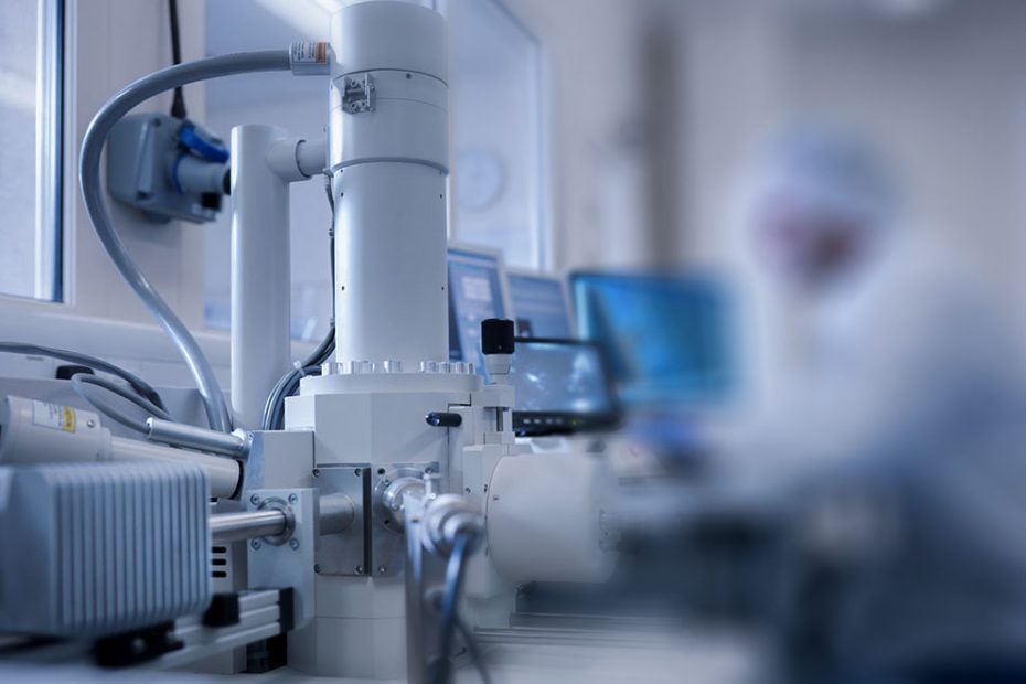
With the rapid development of scientific and technological progress, the magnification of the electron microscope has increased from a dozen times the first electron microscope to a million times today. Therefore, in the biomedical engineering industry, the electron microscope with excellent performance is used to observe various The ultrastructure of normal cell structure and pathology, such as endoplasmic reticulum, membrane protein, Golgi apparatus, lysosome, cytoskeleton apparatus, etc., of great help to discover the source of disease and treat diseases. According to the scientific research on the structure of the cell membrane and the relationship between the structures, it is also possible to scientifically study the laws of cell metabolism such as cell communication and transportation, disintegration and division, reproduction and control, and the electron microscope can also combine various sample preparation techniques to observe virus infection Ultrastructures such as bacteria, chlamydia, and molecular chaperones are irreplaceable special tools for contemporary biomedical engineering research.
(1) Applications in cell biology and molecular biology
Electron microscopes only have very high resolution and magnification. People have been able to observe and study the ultrastructure of subcells, such as the structure of cell membrane, endoplasmic reticulum, cytoskeleton, organelles, etc., and can link the morphological structure with physiological functions. Conduct dynamic studies. A large number of experimental facts of ultrastructural variation of tissues, cells and microorganisms under pathological conditions have greatly enriched the content of cell biology, but have entered the combination of basic medicine and clinical medicine. For example, using the cryo-etching method to observe the inner and outer surfaces of the cell membrane reveals many new phenomena and life science facts that have not been seen in the past.
Ultrahigh-voltage electron microscopy holds promise for direct observation of the living state of living specimens. Electron microscope technology is widely used in the observation and research of the structure of chromosomes and biological macromolecules and provides a powerful tool for morphological research for the development of molecular genetics and biological genetic engineering. At present, it is mainly used in the research of protein, nucleic acid, amino acid series, and gene fragments of transcription and translation.
(2) Applications in Anatomy
At present, electron microscopes can be used to observe and study all human tissues and organs, to observe the fine structure of blood vessels, to study the morphological characteristics of the spatial distribution of microvessels in various tissues and organs, to see the ultrastructure of the bone tissue surface, and The ultrastructure of bone cells and the deposition process of calcium salts in the bone matrix between collagen fibers can be seen.
The application of electron microscopy in anatomy has enabled the understanding of human tissue structure to enter the ultrastructural level and promoted the in-depth development of anatomy. Electron microscopy not only contributes to the morphological study of nerve fibers but also contributes to the development of neurobiology.
(3) Application in virus research
Viruses are the smallest state of life known to humans, and the electron microscope is the only tool for direct observation of them. The discovery of many viruses relies in part on the application of electron microscopy. The study of virus morphology, development and effect on target cells by electron microscope technology provides morphological data for the etiology analysis and prevention of viral diseases.
For viruses that do not cause obvious changes in cells, such as rubella virus and rhinovirus, electron microscopy is a reliable means of identification and diagnosis.
(4) Application in clinical testing
With the research and development of ultrastructural diagnostics, electron microscopy can provide valuable information for clinical clinics of various difficult diseases in blood diseases, tumors, liver and gallbladder, digestion, urinary, skin, etc. For example, auxiliary diagnosis of squamous cell carcinoma, immune nephropathy, various leukemias, etc. Because the ultrastructural lesions of various kidney diseases have their own characteristics, the standard renal biopsy by electron microscope observation is helpful for early diagnosis. Glomerular diseases can be classified according to changes in the glomerular basement membrane in combination with other ultrastructural lesions. According to the characteristics, the differential diagnosis of sclerosing nephritis, congenital nephritis and certain immune nephropathy, etc., can also be used to assist in the diagnosis and identification of some viruses that are difficult to culture in clinical tests, such as hepatitis virus, rotavirus, rubella virus, etc.
(5) Application in ultramicro pathology
At present, many aspects are studied: the changes of nucleus and nucleolus under the action of viruses, carcinogens or poisons; the pathological changes of mitochondria, endoplasmic reticulum, Golgi complex and other organelles caused by ischemia and hypoxia. With the help of an electron microscope, we can also observe the expansion and rupture of the endoplasmic reticulum caused by chemical carcinogens, ischemia and hypoxia, and the degranulation of polyribosomes, which affects the synthesis of proteins. Scanning electron microscopy can be used to find that after Plasmodium invades the human body, it can first attach to the surface of red blood cells like an amoeba, and then enter the red blood cells, causing red blood cell deformation and other pathological processes; Escherichia coli appears on the surface after being treated with kanamycin Such granular drop and so on. These ultramicro pathological or pharmacological observations have certain clinical significance.
Summary
It should be pointed out that the application of electron microscopy in biomedicine also has great limitations. For example, live specimens cannot be observed, and specimens must be made into slices with a thickness of less than 0.1um. It is difficult to detect three-dimensional structures. One of the conditions for electron beam imaging is that there are a certain amount of heavy atoms that can scatter electrons in specimens, while in biological tissues Mainly light atoms such as C, H, U, N. These Achilles’ heels are not only unfavorable factors for the application of electron microscopy in the field of biology, but also important factors that promote the development of microscopic technology.