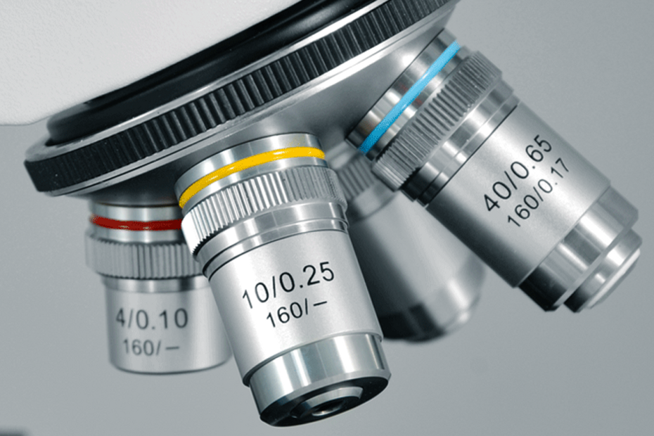
Microscope optical components – objective lens
An objective lens is a lens group composed of several lenses. The purpose of combined use is to overcome the imaging defects of a single lens and improve the optical quality of the objective lens. The magnifying effect of a microscope mainly depends on the objective lens. The quality of the objective lens directly affects the quality of the microscope image. It is the main component that determines the resolution and image clarity of the microscope, so the correction of the objective lens is very important.
Since the objective lens is responsible for the first image formation and plays a central role in determining image quality, it may be called the most important part of an optical microscope.
But with so many types of microscope objectives available, choosing the right one for your application can be a challenge.
In today’s article, we’ve put together a series of questions to help you make the trade-offs and choose the right objective.
01 What is the size of your specimen?
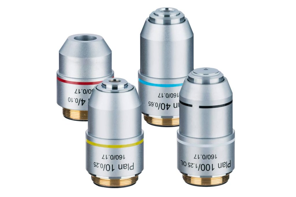
Microscope objectives have a magnification range from 1.25x to 150x. This is the first parameter to consider when choosing the best objective for your application. This parameter, combined with the eyepiece magnification, determines the overall magnification of the microscope.
02 What is the smallest feature in the specimen you want to observe?
The second important parameter of a microscope objective is the numerical aperture (NA). Numerical aperture is a measure of its ability to gather light. This is an important factor in determining resolution, depth of focus, and image brightness. Objectives with larger numerical apertures focus a wider range of light, resulting in brighter, higher-resolution images.
The numerical aperture is also important for observing very fine structures or detecting weak signals during fluorescence observations. Numerical aperture needs to be considered when determining which microscope objective can resolve the smallest features of a specimen. When weighing the pros and cons of your options, keep in mind that numerical aperture ranges from 0.04 to 1.7.
03 What are your image field of view and depth of field requirements?
Field of view and depth of field are two important characteristics of microscope objectives.
Field number or field diameter is the diameter of the field of view in an optical microscope. It is in millimeters and measured at the intermediate image plane. Keep in mind that modern plan apochromats and other specialized plan objectives typically have a usable field of view of 22 to 26.5 mm (or more when combined with wide-angle eyepieces).
The depth of field of an objective lens is the axial focus range of the objective lens without significant changes in image sharpness. This value varies greatly from low to high numerical aperture objectives; it generally decreases as numerical aperture increases.
04 What are your resolution requirements?
The resolution of a microscope objective determines the minimum distance between two observable objects. It is directly proportional to the irradiation wavelength of light and inversely proportional to the numerical aperture.
The higher the numerical aperture, the smaller the distance between two objects. As mentioned previously, choosing an appropriate numerical aperture is critical to determining the resolution of a microscopy system.
05 What kind of working distance do you need?
Working distance (WD) is the distance from the front lens of the objective to the closest surface of the coverslip when the specimen is in focus. Working distance is inversely proportional to numerical aperture, meaning that objectives with higher numerical apertures generally have shorter working distances.
If your application does not require a coverslip and requires a longer distance between the focal plane and the end of the objective (i.e. observing samples with irregular topography, fine structures, or that are mechanically constrained by the overall optical assembly).
06 If you need to observe a fluorescent sample, what is the brightness of the fluorescent signal?
If you are observing a sample with a weak fluorescence signal, it is recommended that you use a high numerical aperture objective that can collect more light.
07 Are you using multi-channel or single-channel fluorescence imaging?
To compensate for chromatic aberration correction, you can use different types of objectives: achromatic objectives, semi-apochromatic objectives, and apochromatic. Achromatic objectives have the lowest correction performance, semi-apochromatic objectives (or fluorite) provide additional spherical correction, and apochromatic objectives provide the highest level of chromatic aberration correction.
If your application requires multi-channel fluorescence, then we recommend using apochromatic objectives.
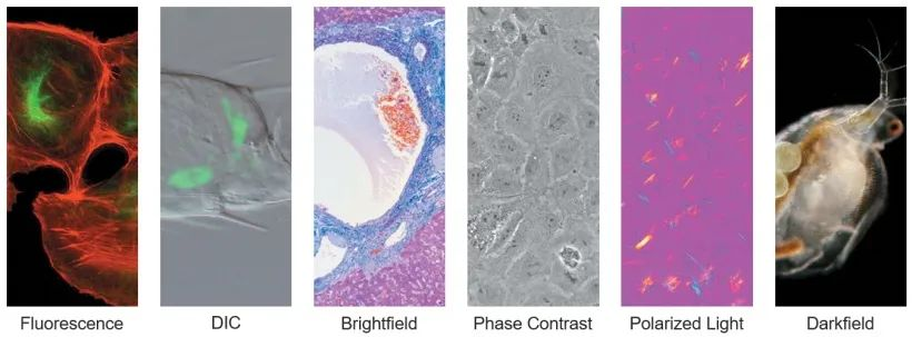
08 Which observation method do you use?
When observing samples that are too thick, too thin, or birefringent, researchers will use other observation methods besides brightfield. Commonly used observation methods are darkfield, differential interference contrast (DIC), phase contrast, and polarized light.
09 What is the medium used for your samples?
Many microscope objectives are designed to image specimens using air as the medium, while others use immersion media with higher refractive index to achieve higher numerical aperture and resolution.
For example, using immersion oil instead of air as the imaging medium can increase resolution by approximately 1.5 times.
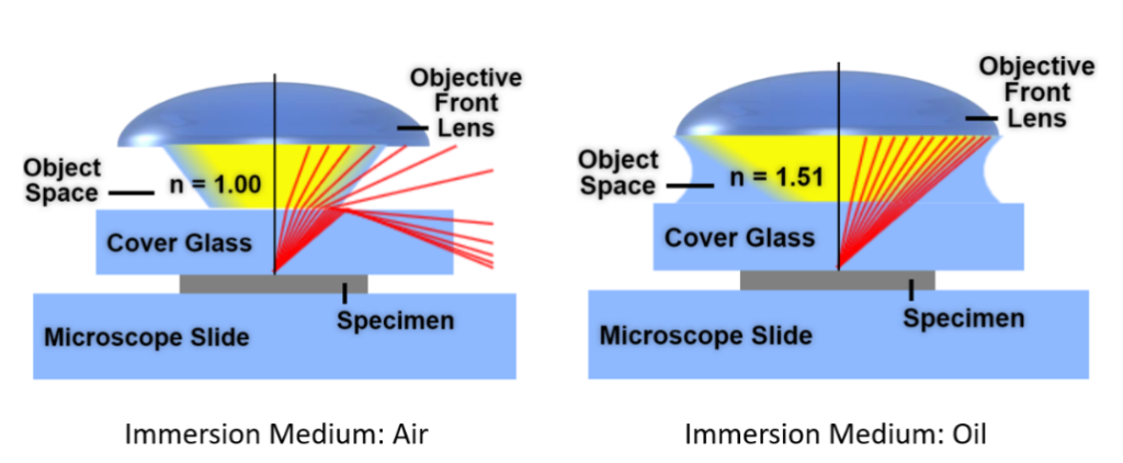
The most common immersion media are air, water, oil, and silicone oil. Choosing the right objective for your media can improve image quality.
10 Are you using a high-end microscope system?
Provides specialized objectives for advanced optical systems such as confocal, spinning disk confocal, multiphoton excitation, and total internal reflection fluorescence (TIRF) microscopy. If you are using this type of system, selecting the correct objective for the application is critical.