Since the first commercial scanning electron microscope came out in 1965, after more than 40 years of continuous improvement, the resolution of the scanning electron microscope has increased from the first 25nm to the current 0.01nm, and most scanning electron microscopes can be used with X-ray spectrometers, The combination of X-ray energy spectrometer and others has become a multifunctional electron microscopic instrument capable of comprehensive analysis of the surface microcosm.
In the field of materials, scanning electron microscopy plays an extremely important role and is widely used in the research of the morphology and structure of various materials, interface conditions, damage mechanisms, and material performance prediction. The scanning electron microscope can be used to directly study crystal defects and their generation process, to observe the aggregation mode of atoms inside the metal material and their real boundaries, to observe the way of boundary movement under different conditions, and to check the crystal defects caused by surface machining. damage and radiation damage.
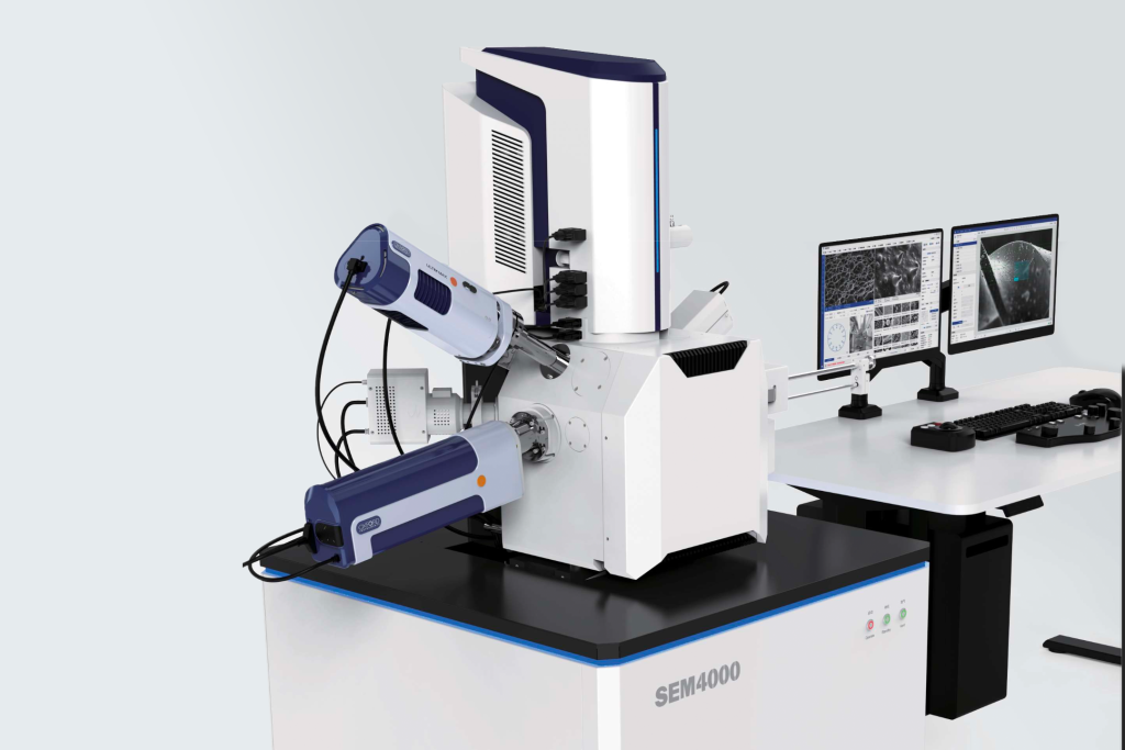
Advantages of Scanning Electron Microscopy (SEM):
- High magnification, continuously adjustable between 200,000 and 200,000 times;
- It has a large depth of field, a large field of view, and three-dimensional imaging, which can directly observe the fine structure of the uneven surface of various samples;
- Sample preparation is simple.
The current scanning electron microscope is equipped with an X-ray energy spectrometer device, which can simultaneously observe the microstructure and appearance and analyze the composition of the micro-area, so it is a very useful scientific research instrument today.
Four elements that affect SEM
1. Resolution
The main factors affecting the resolving power of SEM are:
A. The diameter of the incident electron beam spot: it is the limit of the resolving power of the scanning electron microscope. Generally, the minimum beam spot diameter of the hot cathode electron gun can be reduced to 6nm, and the field emission electron gun can make the beam spot diameter smaller than 3nm.
B. Expansion effect of the incident electron beam in the sample: the degree of diffusion depends on the energy of the incident beam electrons and the atomic number of the sample. The higher the energy of the incident beam, the smaller the atomic number of the sample, the larger the volume of the electron beam, and the area where the signal is generated increases with the diffusion of the electron beam, thereby reducing the resolution.
C. Imaging method and modulation signal used: when the secondary electron is used as the modulation signal, due to its low energy (less than 50 eV), the mean free path is short (about 10-100nm), only in the depth range of 50-100nm in the surface layer Only the secondary electrons in the sample can escape from the sample surface, and the number of scattering is very limited and basically does not expand laterally.
Therefore, the resolution of the secondary electron image is approximately equal to the diameter of the beam spot. When the backscattered electrons are used as the modulation signal, the energy of the backscattered electrons is relatively high and the penetrating ability is strong, so they can escape from a deeper area in the sample (about 30% of the effective depth).
In this depth range, the incident electrons have a fairly wide lateral expansion, so the resolution of the backscattered electron image is lower than that of the secondary electron image, generally around 500-2000nm. If other operation modes such as absorption of electrons, X-rays, cathode fluorescence, beam-induced conductance or potential are used as modulation signals, since the signal comes from the entire electron beam scattering area, the resolution of the obtained scanning image is relatively low, generally at 1000nm or 10000nm The above varies.
2. Magnification
The magnification of the scanning electron microscope can be expressed as
M=Ac/As
In the formula, Ac—the side length of the image on the fluorescent screen;
As—the scanning amplitude of the electron beam on the sample.
Generally, Ac is fixed (usually 100 mm), and the magnification can be changed by changing As. At present, the magnification of most commercial scanning electron microscopes is 20 to 20,000 times, which is between the optical microscope and the transmission electron microscope, that is, the scanning electron microscope makes up for the gap in the magnification of the optical microscope and the transmission electron microscope.
3. Depth of field
Depth of field refers to a distance range before and after the focal point, and the image formed by all object points in this range meets the resolution requirements and can form a clear image; that is, the depth of field is the distance range that can be seen clearly. The depth of field of a scanning electron microscope is 10 times greater than that of a transmission electron microscope and hundreds of times greater than that of an optical microscope. Due to the large depth of field of the image, the obtained scanned electronic image has a three-dimensional effect. The depth of field of the electron beam depends on the critical resolution d0 and the incident half-angle αc of the electron beam.
Among them, the critical resolving power is related to the magnification, because the resolving power of the human eye is about 0.2 mm. After zooming in, in order to make the object clear, the resolution of the electron beam must be higher than the critical resolution d0, and the incident electron beam The angle can be adjusted by changing the aperture size and working distance, and a small incident electron angle can be obtained with a small aperture and a large working distance.
4. Contrast
Including surface topography contrast and atomic number contrast. Surface topography contrast is caused by the unevenness of the sample surface. Atomic number contrast refers to the backscattered electrons, absorbed electrons, and X-rays generated when the scanning electron beam is incident on the test screen, and is quite sensitive to the difference in atomic number in the micro-area. The larger the atomic number, the brighter the image. Secondary electrons are less affected by atomic number. The average atomic number of each component in the polymer has little difference; so only some special polymer multiphase systems can use this contrast imaging.