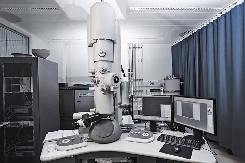
1. What is cryo-electron tomography?
Using cryo-electron tomography (also known as electron tomography), protein-protein interactions can be analyzed at three-dimensional molecular resolution in both native and functional states.
As the sample is tilted in a series of controlled positions, it is imaged as a series of two-dimensional images. The resulting image “slices” can be combined to produce a 3D reconstruction of the sample.
Cryo-electron tomography (Cryo-ET) is an imaging technique used to obtain high-resolution three-dimensional reconstructions of biomolecules.
In cryo-electron tomography, the vitrified sample is imaged in a transmission electron microscope at an inclination of approximately -60° to +60°. This produces a series of 2D images, known as tomograms, that can be combined to reconstruct a three-dimensional image.
Powerful technical functions, enough to analyze the 3D structure of intracellular organelles and protein complexes in a near-native state in the cellular environment.
Because the location and structure of a biomolecule are critical to understanding its cellular function, resolving structure in a cellular context is a breakthrough in both structural and cell biology.
2. What is the general process of cryo-EM structure analysis?
The general process of analyzing protein structure by cryo-electron microscopy is as follows:
protein expression and purification
Negative stain sample preparation: about 2 hours to complete
Data collection of negatively stained samples: about 8 hours to complete
Preparation of frozen samples: about 4 hours to complete
Data collection for frozen samples: 48-120 hours to complete
3D structure reconstruction.
3. What are the requirements for the sample to analyze the structure of the cryo-electron microscope? Where is the difficulty?
Cryo-electron microscopy analysis of protein structure requirements for proteins:
Molecular weight: Generally, the molecular weight of the sample is required to be above 200kD
Buffer: The buffer should not contain polysaccharides, DMSO, glycerol, and other organic substances, which will reduce the contrast of the sample and make it difficult to obtain a high-resolution three-dimensional structure. Generally, the buffer is 20 mM Hepes and 150 mM NaCl.
Concentration: Generally speaking, the concentration of soluble protein should be around 1mg/ml, and the concentration of membrane protein should be around 5mg/ml.
Volume: 20ul is enough (the premise is that the protein concentration needs to reach the standard, and make a sample of about 3ul).
Uniformity: Molecular sieve behaves as a single peak, and the uniformity is greater than 90%.
Explanation: The main risks of cryo-electron microscope analysis structure are:
A. The sample is very unstable. When the sample is sent, it has been degraded or aggregated, and subsequent processing cannot be performed;
B. During the process of freezing samples, the samples may be frozen and shattered, making subsequent processing impossible;
C. The purity of the sample may be good, but the uniformity is poor, making it difficult to obtain the high-resolution structure of the sample;
D. The area we care about may have strong flexibility in the entire complex, so after two-dimensional or three-dimensional averaging, the resolution of the area we care about will be relatively poor or even invisible, which cannot meet our expectations;
E. Some ligands, such as prodrug molecules, have too small molecular weight to be observed in the density map of the electron microscope;
F. For suitable samples, we need to optimize many parameters for frozen samples, including optimization of sample concentration, optimization of blot time, optimization of temperature, optimization of grid specifications (copper grid or gold grid), and a series of optimization conditions.
Therefore, cryo-electron microscopy sample preparation requires rich experience and sufficient machine time support, and the success rate may vary greatly among different experimenters.
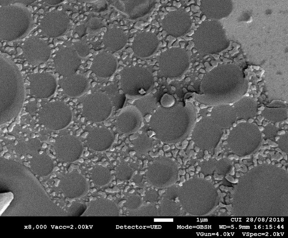
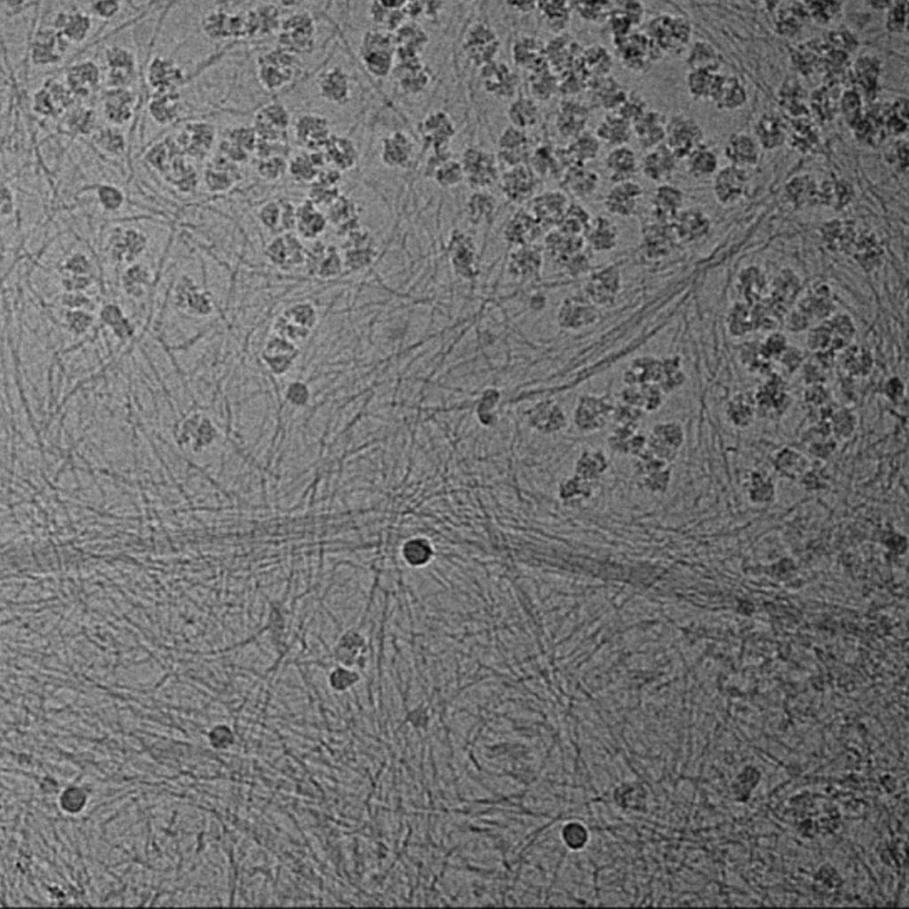
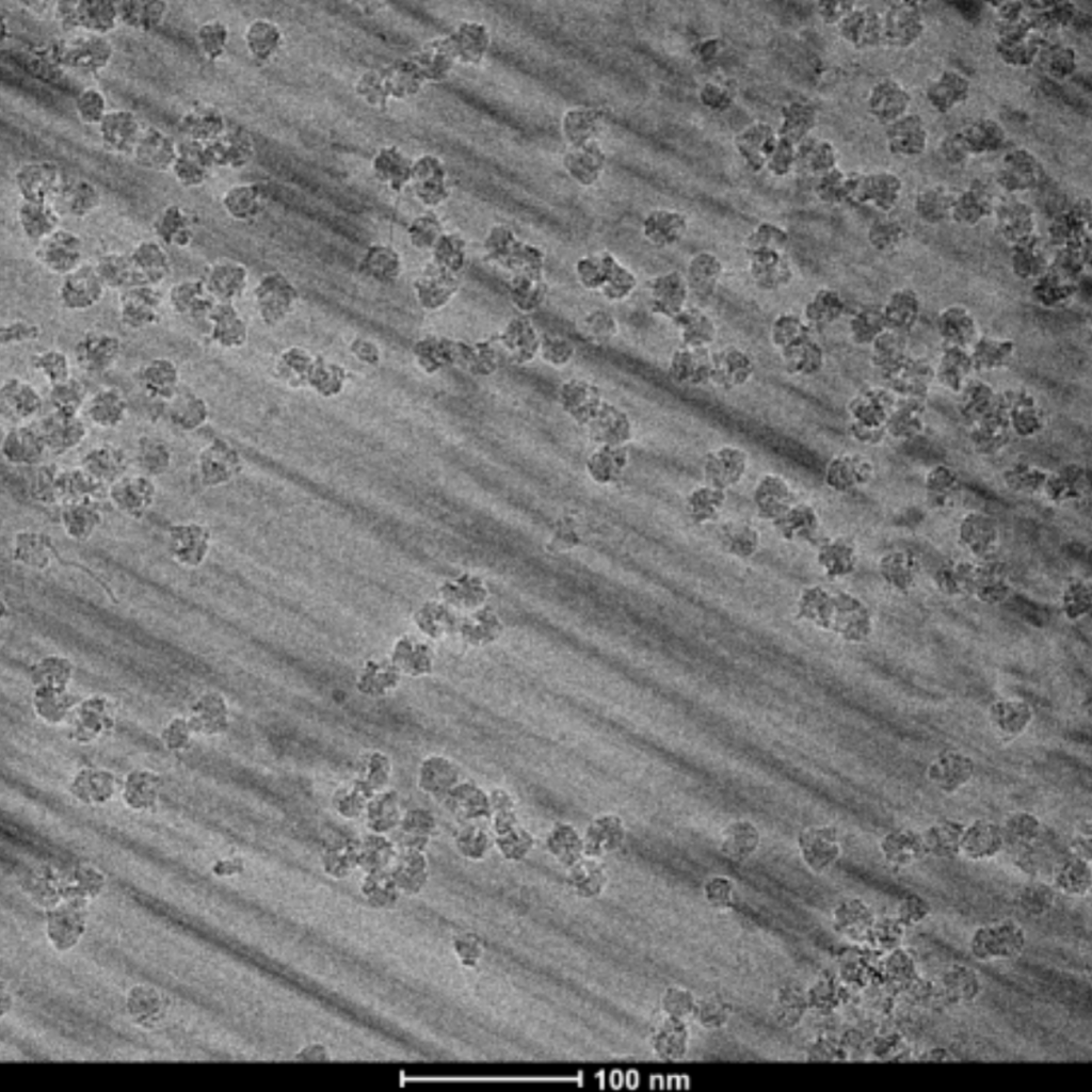
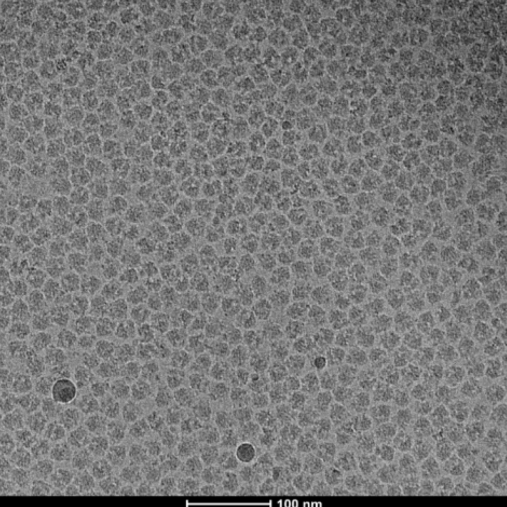
Examples
4. What research fields is cryo-electron tomography used in?
Cryo-electron tomography was originally used primarily in structural biology, but due to recent technological advances, it has become a powerful tool widely used in microbiology, neurobiology, immunology, cell biology, developmental biology, molecular biology, and other broad research fields.
Microbiology researchers use cryo-electron tomography to visualize viral infection in bacteria. For example, by using cryo-electron tomography, researchers can reconstruct the structure of viruses at different stages of infection. In another study, they used cryo-electron tomography to show that viruses create nucleus-like structures in bacteria to protect themselves from defense mechanisms.
Cryo-electron tomography is used in neurobiology to visualize protein aggregation that leads to neurodegenerative diseases. Traditional neuronal cell sample preparation methods are not capable of high-resolution imaging, but by using cryo-electron tomography, the researchers were able to visualize aggregates associated with Huntington’s disease and lateral sclerosis.
In immunology, the researchers used cryo-electron tomography to study the structure of HIV-1 particles bound to HeLa cells, revealing their interactions.
In cell biology, cryo-electron tomography has been used to elucidate connections between different intracellular membrane systems through membrane contact sites. High-resolution 3D reconstructions from cryo-electron tomography allow the study of molecules involved in these rare contacts between different organelles.
To date, cryo-electron tomography has mainly been performed using cultured cells or single-celled organisms. In order to use cryo-electron tomography for the developmental biology of multicellular organisms, new protocols have now been developed that allow classical model organisms such as C. Elegans and D. Melanogaster.
Recent research in the field of molecular biology has revealed important structural details of the mechanism of action of Cas9 using cryo-EM purified proteins. Taking this research a step further by capturing CRISPR-Cas9 in situ using cryo-electron tomography will greatly help optimize this famous gene editing system.