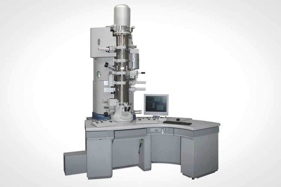
Scanning transmission electron microscopy is a development of transmission electron microscopy. It is a powerful micro-area material characterization technique, which can obtain various information such as sample morphology, composition, and microstructure.
The development of spherical aberration correction technology has advanced its resolution to the sub-angstrom level, making it possible to study the atomic structure and electronic structure of materials at the atomic level.
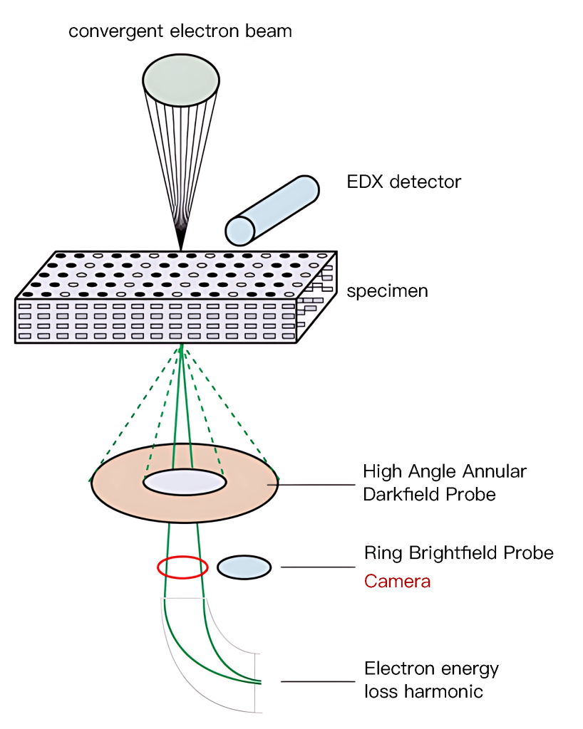
The scanning coil of the scanning transmission electron microscope forces the electron probe to scan on the thin film sample. The difference from the scanning electron microscope is that the detector is placed under the sample, and the detector accepts the transmitted electron beam or elastically scattered electron beam. Finally, the bright field image and dark field image of the scanning transmission electron microscope corresponding to the conventional transmission electron microscope are displayed on the fluorescent screen.
Instrument Features:
1. Scanning transmission electron microscopy has high technical requirements, a very high degree of vacuum, and the electronic system is more complicated than TEM and SEM.
2. The acceleration voltage is low, which can significantly reduce the damage of the electron beam to the sample and can greatly improve the contrast of the image, especially suitable for the transmission analysis of soft material samples such as organic polymers and biology.
3. It can observe thicker samples and samples with low contrast.
4. STEM can perform energy-dispersive X-ray spectroscopy (EDS) or electron energy-loss spectroscopy (EELS) to analyze the elemental composition of the sample.
5. The strong excitation of the objective lens in the scanning transmission mode can realize micro-diffraction.
6. STEM can operate in various imaging modes, such as bright-field imaging, dark-field imaging, and high-angle annular dark-field (HAADF) imaging, allowing different contrast mechanisms for specific applications.
7. When using the scanning transmission electron microscope mode of the scanning electron microscope to observe biological samples, the samples can be directly observed without staining to obtain images with higher contrast.
Maintenance:
1. Remove the dust cover and turn on the power.
2. Put the sample on the spacer of the stage, and adjust the coarse/fine adjustment knob to adjust the focus until the observed image is clear.
3. Adjust the position of the stage, find the field of view to be observed, and conduct a metallographic analysis.
4. When adjusting the focus of the scanning electron microscope, be careful not to let the objective lens touch the sample, so as not to scratch the objective lens.
5. Do not switch the objective lens when the center of the round hole of the stage spacer is far away from the center of the objective lens, so as not to scratch the objective lens.
6. Do not adjust the brightness suddenly and suddenly, and do not make it too bright, which will affect the service life of the bulb and also damage the eyesight.
7. For all (function) switching, the action should be light and in place.
8. When shutting down, adjust the brightness to the minimum.
9. Non-professionals should not adjust the metallographic microscope lighting system (filament position lamp), so as not to affect the imaging quality.
10. When replacing the halogen lamp, pay attention to high temperature to avoid burns; be careful not to directly touch the glass body of the halogen lamp with your hands.