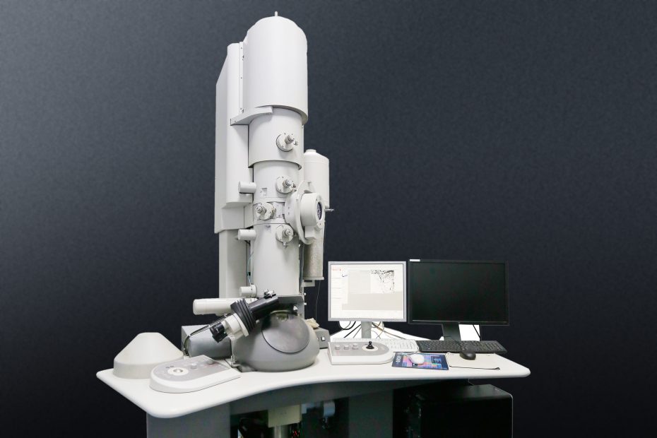
Transmission electron microscopy (hereinafter referred to as TEM) is a technique used to observe very small sample features. The technique uses a beam of accelerated electrons, which travels through very thin samples, allowing scientists to observe features such as structure and morphology.
In 1932, German scientists Max Knoll and Ernst Ruska invented the transmission electron microscope with an electron beam as the light source. The wavelength of the electron beam is much shorter than that of visible light and ultraviolet light, and the wavelength of the electron beam is inversely proportional to the square root of the voltage of the emitted electron beam. That is to say, the higher the voltage, the shorter the wavelength. TEM can see microstructures smaller than 0.2um that cannot be seen clearly under an optical microscope. These structures are called sub microstructures or ultrastructures. After years of development, it has become a common technique for observing micro- and nano-particles in the world of science and engineering.
Advantages of TEM
Transmission electron microscopy can tell us about the structure, crystallization, morphology and stress of a substance while scanning electron microscopy can only provide information about the morphology of a sample. However, TEM requires very thin samples that are translucent to electrons, which can mean that sample preparation takes longer.
The six main system components of TEM
The system components of the transmission electron microscope are mainly composed of the electron gun, condenser lens, sample chamber, objective lens, intermediate mirror and transmission mirror.
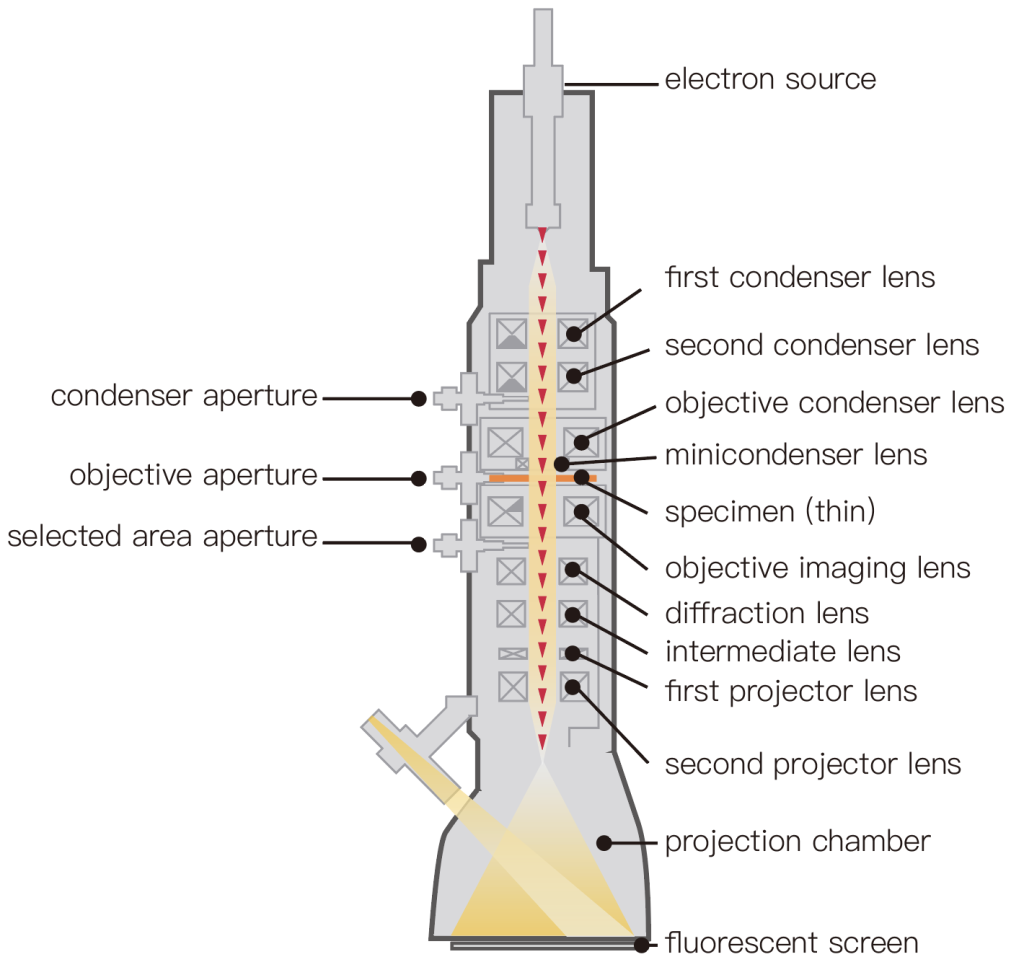
- Electron gun: emits electrons and consists of a cathode, a grid, and an anode. The electrons emitted by the cathode tube form a ray beam through the small hole on the grid, and after being accelerated by the anode voltage, it shoots to the condenser lens, which accelerates and pressurizes the electron beam.
- Condenser: Focuses the electron beam and can be used to control the illumination intensity and aperture angle.
- Sample room: place the sample to be observed, and is equipped with a tilting table to change the angle of the sample, as well as equipped with heating, cooling and other equipment.
- Objective lens: It is a short-distance lens with high magnification, and its function is to magnify the electronic image. The objective lens is the key to determining the resolving power and imaging quality of the transmission electron microscope.
- Intermediate mirror: It is a weak lens with variable magnification, and its function is to re-magnify the electronic image. By adjusting the current of the intermediate mirror, the image or electron diffraction pattern of the object can be selected for amplification.
- Transmission mirror: It is a high-power strong lens, which is used to magnify the intermediate image and image it on the fluorescent screen.
In addition, there is a secondary vacuum pump to evacuate the sample chamber and a camera device to record images.
How TEM Works
In a transmission electron microscope, an electron gun emits a beam of electrons. The gun uses electromagnetic coils and voltages of up to millions of volts to accelerate electrons to extremely high speeds.
The electron beam is focused into a fine beam by a condenser lens with a high aperture to eliminate high-angle electrons. At top speed, the electrons zip through the ultra-thin sample, with portions of the electron beam transported depending on how transparent the sample is to electrons.
The objective lens focuses the portion of the beam emitted from the sample into an image. Another component of the TEM is the vacuum system, which is essential to ensure that electrons do not collide with gas atoms.
Sub-vacuum is first achieved using a rotary or diaphragm pump, which can provide a pressure low enough for a diffusion pump to operate, and then reach a vacuum level high enough to operate. High-pressure TEMS requires particularly high vacuum levels, and a third vacuum system can be used.
The images produced by the TEM are called photomicrographs and are viewed by projecting onto a phosphorescent screen. When illuminated by an electron beam, the screen emits photons. A film camera located below the screen can be used to capture the image, or a charge-coupled device (CCD) camera can be used for digital capture.
Applications of TEM
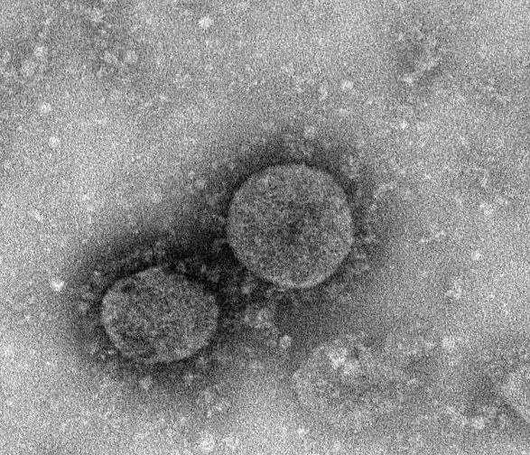
The technology is used in various industries, from medical research to study viruses and bacteria, to forensics, gemmology and materials science. Transmission electron microscopy is widely used in materials science and biology. Because electrons are easy to scatter or absorb by objects, the penetrating power is low, and the density and thickness of the sample will affect the final imaging quality. Thinner ultrathin sections must be prepared, usually 50-100nm. Therefore, the sample must be processed very thinly when observed with a transmission electron microscope. Commonly used methods are the ultrathin section method, frozen ultrathin section method, freeze etching method, freeze-fracture method, etc. For liquid samples, it is usually observed on a pretreated copper grid.