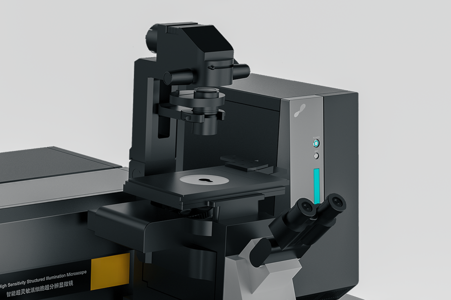
Ultra-high-resolution fluorescence microscopy is constantly changing our understanding of the internal structure and operation of cells. However, at this stage, microscope technology still has various shortcomings. If people hope that microscopes can play an important role in the field of biological research, they must be improved and improved.
1. The emergence of optical microscopes and their impact
Microscopes have always been an indispensable tool for biologists to research and explore life’s mysteries. It is precisely because of this great invention of Leeuwenhoek (Dutch naturalist and microscope creator, 1632-1723) and the continuous improvement and development of microscope technology by his successors that people can understand the intricate subcellular organelles inside cells. We have a preliminary understanding of the shape of the structure.
Since then, researchers have never stopped pursuing microscopy technology. They always hope to obtain microscopes with higher resolution to observe the finer structures inside cells better. Recently, super-resolution imaging (SR imaging) has finally made it possible to observe and study at the single-molecule level.
2. The development process of ultra-high-resolution imaging technology
The process of reaching today’s level of ultra-high-resolution imaging technology has involved the hard work of many researchers and also faced many problems that need to be solved.
Among the above optical ultra-high-resolution imaging technologies, two technologies – stimulated emission depletion microscopy (STED) and saturated structured illumination microscopy (SSIM) – have attracted the most attention.
Recently, photosensitive localization microscopy (PALM) and stochastic optical reconstruction microscopy (STORM) based on probe ultra-high-resolution imaging technology, as well as fluorescence photosensitive localization microscopy (FPALM) that relies on the random activation properties of fluorophores have achieved success.
Through probe-based ultra-high-resolution imaging technology, multiple raw images can be obtained. In each original image, only a part of the fluorescently labeled molecules in the cell can emit fluorescence, that is, these fluorescent molecules are in an alternating state of activation and deactivation, and only part of the molecules can be observed and imaged each time. And because the molecules that emit fluorescence each time are dispersed relatively sparsely, they will not be affected by each other, thus avoiding the problem of being unable to distinguish adjacent molecules due to their fluorescence. Finally, these original images are superimposed and overlapped to obtain the final high-resolution image. In this way, the resolution of structures that were previously too dense to be imaged due to fluorescent dots can be reached to the nanometer level, and the density of imaged molecules is also quite high, which can reach 105 molecules/μm2.
This resolution means for biologists that it is now possible to observe structures and dynamic processes within cells at the molecular level.
Although microscopy technology has advanced to such great heights, it is still just a tool used by biologists in research. Therefore, it is necessary to cross-reference the images obtained by the microscope with other test results to obtain accurate results. People need to recognize the advantages and disadvantages of ultra-high-resolution microscopy and develop standardized operating specifications for operating and judging ultra-high-resolution imaging technology images. Only in this way can the role of ultra-high-resolution microscopy be maximized.
Nowadays, since people only have a conceptual understanding of the organizational structure of various components in cells and their dynamic changes, using microscopes to observe these structures and processes at the nanometer level truly can allow people to discover many things in the past. Things you don’t understand. For example, when people discovered through electron microscopy that the cytoskeleton is composed of a large number of filamentous mesh-like tissues, some people were skeptical about this phenomenon. Cell biologists who believe that the cytoskeleton is a structure used to dilute the biochemical soup inside the cell call this observation a rigid artifact.
Unless the latest ultra-high-resolution microscopy images or other experimental results can prove that the cytoskeleton is composed of a large number of filamentous mesh-like tissues, some people will still hold the above-mentioned skeptical view. However, other biochemical test results have confirmed that the early electron microscope observations were correct. Of course, the emerging ultra-high-resolution imaging technology also needs to be supported by the results of other traditional biochemical tests to be valuable, and it also needs the assistance of electron microscopy. Because electron microscopy can provide nanometer-scale observations, it is most valuable for corroborating ultra-high-resolution microscopy observations with the same resolution.
In the future, as everyone gradually understands, accepts, and widely uses ultra-high-resolution microscopes, they need to pay attention to various problems that will arise. The following table lists some of the shortcomings related to the use of ultra-high-resolution microscopes and their current problems. Solution.
In recent years, there have been very strict regulations on how to process images. But so far, there is no standard operating protocol for how to process ultra-high-resolution imaging technology images. In particular, it should be pointed out that PALM and STORM data have more in common with graphs than images on some important factors. The uncertainty and density of a molecule are represented by color in an ultra-high-resolution imaging image, marking any location within the cell where the molecule might appear. Only after the labeled molecule is judged to be a single molecule according to certain standards (the number of photons emitted) and its positioning is accurate, it will be displayed. Such normalization of the acquired images must be performed before the results can be analyzed. Likewise, experimental data also need to be standardized. To improve the resolution, not only the molecules must be positioned and distributed well, but also the number of molecules must be large enough to meet the requirements of the Nyquist criterion, that is, the average distance between molecules must be less than half the resolution of the microscope. Although the above problems will not affect the application of ultra-high-resolution microscopy, due to the existence of these problems, we should always remind ourselves that we must carefully interpret and analyze the image results of ultra-high-resolution microscopy. Only in this way can we obtain valuable Biological conclusions.
3. Application of ultra-high-resolution fluorescence microscopy in biological research
So far, it’s hard to know which area of biology ultra-high-resolution fluorescence microscopy will bring major changes to, but there are already obvious changes in several areas. These research fields are dynamic and static cell tissue structure research, heterogeneous molecular organization research, and protein dynamic assembly research. These fields all have a common feature, that is, their research focuses on how molecules interact and assemble to form complexes. Therefore, it is of great significance to them to be able to observe these molecules at the nanometer level.
3.1 By observing the combinatorial relationship between proteins, we can understand their functions and lay the foundation for subsequent cell function tests.
Structural biology research has made great progress in this area, and has now discovered the mechanism by which intermolecular interactions of 4-8 nanometers in size assemble into cellular microtubules, myofilaments, and intermediate filaments, which are polymers exceeding 10 microns in size. However, better methods are needed to study complexes such as nuclear pore complexes, centrosomes, centromeres, intermediates, and focal adhesions, which are composed of many different proteins through complex three-dimensional assembly. The goal is to achieve molecular-level resolution so that individual molecules can be observed during the formation of large complexes and thus gain some understanding of their stoichiometry. To obtain more biological information, three-dimensional imaging technology such as ultra-high-resolution microscopy is needed. For example, ultra-high-resolution imaging of living cells can be used to capture the dynamic reconstruction process of the cytoskeleton, etc.
3.2 Ultra-high-resolution imaging helps people better understand the differences between molecules
The classic model of how cell membrane proteins are organized has changed from a randomly distributed liquid mosaic model to a lipid raft model, a caveolae model, or a specialized protein model. This difference is related to different functions of the cell. For example, interactions must occur between the Golgi apparatus, cargo proteins, and Golgi enzyme proteins, but eventually, they will be separated according to their respective functions and play their respective roles. Many experimental methods, such as immunoelectron microscopy and fluorescence resonance energy transfer technology (FRET), have been used to study this membrane inhomogeneity problem. Multicolor PALM technology provides people with a new means to observe the process of membrane protein assembly and organization, and can also quantitatively analyze the spatial distance relationship between different proteins. Because of the single-molecule information provided by PALM, people can clearly understand the spatial relationship between protein molecules, and it is even possible to calculate the possibility of interaction between molecules separated by a certain distance. In addition to studying membrane proteins, this method can also be used to study many non-randomly distributed biological systems, such as studying motor proteins on microtubules.
3.3 Ultra-high-resolution imaging technology can also be used to study the dynamic assembly process of proteins at the single-molecule level
The cell’s response to external stimulation signals begins at the cell membrane, where a dynamic collection of receptor proteins occurs to regulate the cell’s response activity. Enveloped viruses like HIV are also viruses that complete the assembly process of virus particles on the cell membrane and also use the cell’s material transport mechanism. Although the physical model of protein assembly is far from complete, researchers know that the dynamic assembly process of membrane proteins is not uniform, so it is usually difficult to obtain molecular-level information using fluorescence experiments. Similarly, single molecule measurement technology (Single molecule measurements) also has similar limitations, because single molecule measurement technology can only observe a few molecules within the cell, so it lacks overall information. It is therefore difficult to study protein assembly processes dynamically due to lack of spatial resolution. Combining ultra-high-resolution fluorescence imaging technology with live-cell imaging technology and single-molecule tracking technology (sptPALM) can solve this problem. We can accurately see protein clusters in PALM images with the help of molecular density. Statistical and morphological data of protein cluster dynamics can help us understand the mechanism of dynamic protein assembly.
4. Summary
The above only selected three aspects of biological research to illustrate the use of ultra-high-resolution imaging technology, but this has already demonstrated the use of ultra-high-resolution microscopy to confirm the structure of molecules, the most basic materials that make up living organisms. Combination process. Commercial products of STED and PALM are already on the market, marking the advent of the era of ultra-high-resolution microscopy.
We believe that in the hands of creative biologists, ultra-high-resolution microscopes will play their full role and help us discover more mysteries of life.