Electron microscope has three main parameters: resolution, magnification, and depth of field.
Resolution
Resolution is the most important performance index of SEM. For imaging, it refers to the minimum distance between two points that can be resolved; for micro-area component analysis, it refers to the smallest area that can be analyzed.
The resolution of the scanning electron microscope is determined by measuring the minimum distance between two particles (or regions) in the image by dividing the minimum distance measured on the image by the magnification under the condition of known magnification The resulting value is the resolution.
The resolution of the scanning electron microscope is related to the type of detection signal. The following table shows the imaging resolution of the main signal of the scanning electron microscope.
| Signal | Backscattered electrons | Secondary electrons | Characteristic X-rays | Absorbed electrons | Auger electrons |
| Resolution/nm | 50-200 | 5-10 | 100-1000 | 100-1000 | 5-10 |
It can be seen from the data in the table that the resolution of secondary electrons and Auger electrons is not high, while the resolution of microscopic images modulated by characteristic X-rays is the lowest.
The reasons for the difference in resolution caused by different signals can be illustrated in the figure below. After the electron beam enters the surface of the light element sample, a drop-shaped interaction volume will be formed. The incident electron beam will travel within this volume before being absorbed by or scattered out of the sample.

Due to their low energy and short mean free path, secondary electrons, and Auger electrons can only escape in the shallow surface of the sample. In general, the thickness of the sample surface that can excite Auger electrons is about 5- 2nm, and the layer depth for exciting secondary electrons is in the range of 5-10nm.
When the incident electron beam enters the shallow surface, it has not expanded laterally, so the secondary electrons and Auger electrons can only be excited in a cylinder with the same diameter as the incident electron beam spot, because the beam spot diameter is an image The size of the detection unit (image point), so the resolution of these two electrons is equivalent to the diameter of the beam spot.
When the incident electron beam enters the deeper part of the sample, the range of lateral expansion becomes larger, and the energy of the backscattered electrons excited from this range is very high, and they can be ejected from the deeper part of the sample to the surface, and the effect of lateral expansion The volume size is the imaging unit of the backscattered electrons, which greatly reduces its resolution.
The incident electron beam can also excite characteristic X-rays in deeper parts of the sample. Judging from the X-ray action volume in the figure, if X-ray modulation is used for imaging, its resolution is lower than that of backscattered electrons.
It should be noted that when the electron beam is injected into the heavy element sample, the action volume is not in the shape of a drop, but in the shape of a hemisphere. The electron beam expands laterally immediately after entering the surface, so when analyzing heavy elements, it cannot achieve high resolution even if the electron beam’s beam spot is very small.
The resolution of the SEM depends on the diameter of the incident electron beam, the smaller the diameter, the higher the resolution, but the resolution is not directly equal to the diameter of the incident electron beam.
Because the effective excitation range of the electron beam in the sample greatly exceeds the diameter of the incident electron beam.
In addition, the scanning electron microscope’s resolution is affected not only by the diameter of the electron beam and the type of modulation signal, but also by the atomic number of the sample, stray magnetic field, mechanical vibration, and other factors.
Magnification
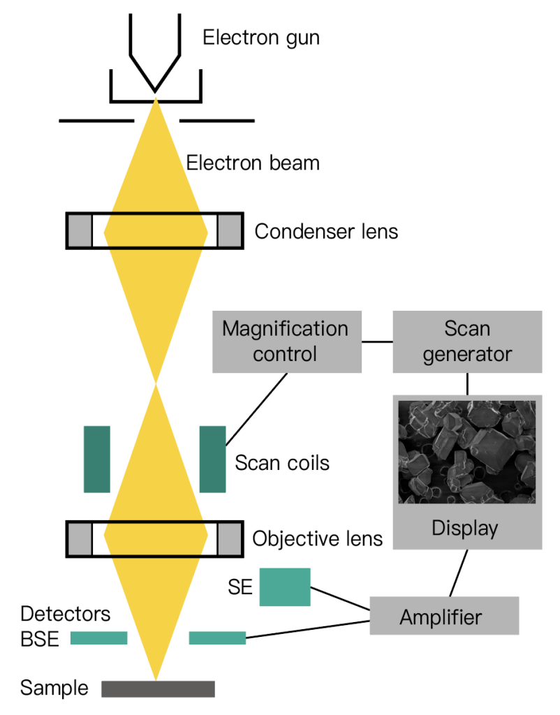
When the incident electron beam is raster scanned if the scanning amplitude of the electron beam on the sample surface is As, and the synchronous scanning amplitude of the cathode ray on the display screen is Ac, then the magnification can be expressed as M=Ac/As.
Since the size of the fluorescent screen is constant, the change in the magnification can only be realized by changing the scanning amplitude As of the electron beam on the sample surface.
The magnification of the current commercial scanning electron microscope can be continuously adjusted from 20 times to 200,000 times.
Depth of Field
Depth of field refers to the ability range of the lens to simultaneously focus and image various parts of the uneven sample, and this range is represented by a certain distance.
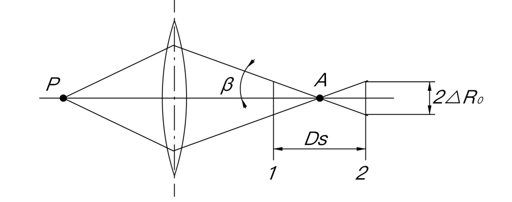
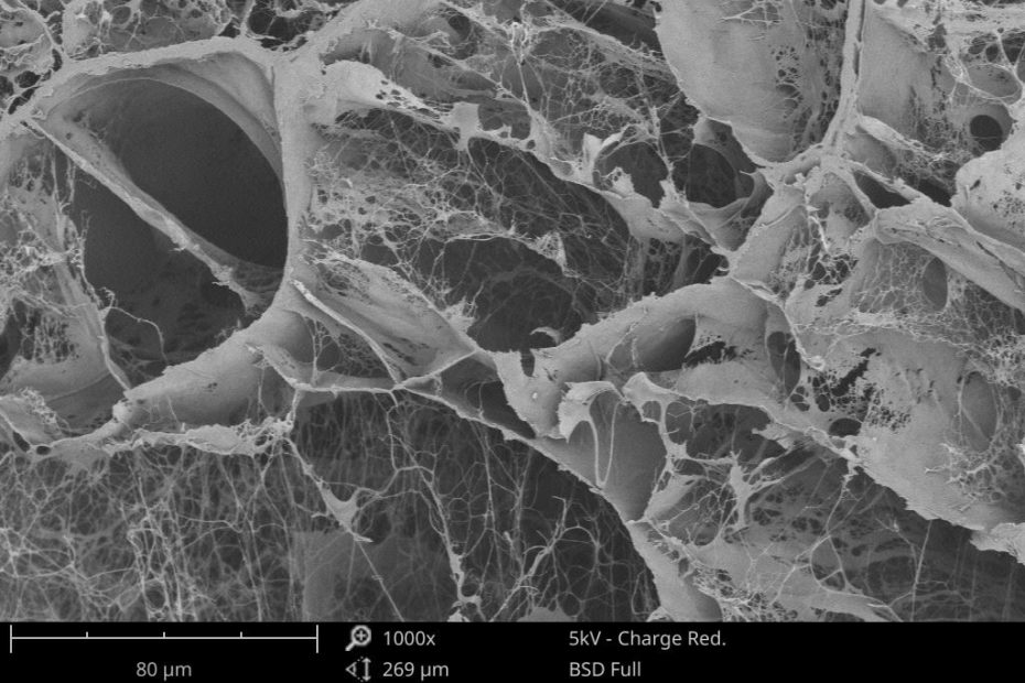
If the depth of field is Ds, as long as the range value of the sample surface is less than Ds, the surface morphology of the sample can be clearly reflected on the fluorescent screen. It is precise because of the large depth of field of the scanning electron microscope that it is especially suitable for the analysis and observation of rough surfaces and fractures; the image is full of three-dimensional, realistic, and easy to identify and explain.