The electron beam acceleration voltage (5kV) used by the low-voltage electron microscope (LVEM) is much lower than that of the large-scale TEM.
A lower accelerating voltage will enhance the strength of the interaction between the electron beam and the sample, thereby improving image contrast and contrast, especially suitable for samples such as polymers and organisms; at the same time, the low-voltage transmission electron microscope has less damage to the sample and has a larger resolution. Low, 1-2nm.
Due to the low voltage, it is possible to integrate TEM, SEM, and STEM in one device. It can provide virology researchers with a variety of imaging modes required for characterization at the same time, and comprehensively analyze the structure and composition of virus samples (Figure 1).
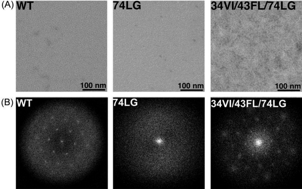
Taking virus detection as an example, since viruses are usually small in size and the main components are mostly carbon-containing light organic elements, such samples are easily penetrated by high-energy electron beams, resulting in low optical contrast. Traditional optical microscopes often difficult to meet the needs of morphological observation, which makes high-resolution transmission electron microscopy an important means of current virological research (Figure 2).
The morphological observation of viruses generally adopts negative staining imaging technology, which requires complex negative staining operations on samples before observation, which takes up a lot of time and may cover up some of the morphological characteristics of viruses, resulting in the use of transmission electron microscopy to observe viruses. The threshold is high.
In order to solve this problem, low-voltage transmission electron microscopy (LVEM) came into being.
LVEM breaks through the minimum acceleration voltage limit of 80kV of the traditional transmission electron microscope. Researchers can observe light biological samples at low pressure without staining, which simplifies the sample preparation process. At the same time, the device can be used under the premise of ensuring high image contrast. Gentle accelerating voltages allow the detection and identification of virus morphology, enabling the identification of viral features that may have previously been masked by stains and negative staining blemishes.
Features of low-voltage compact TEM
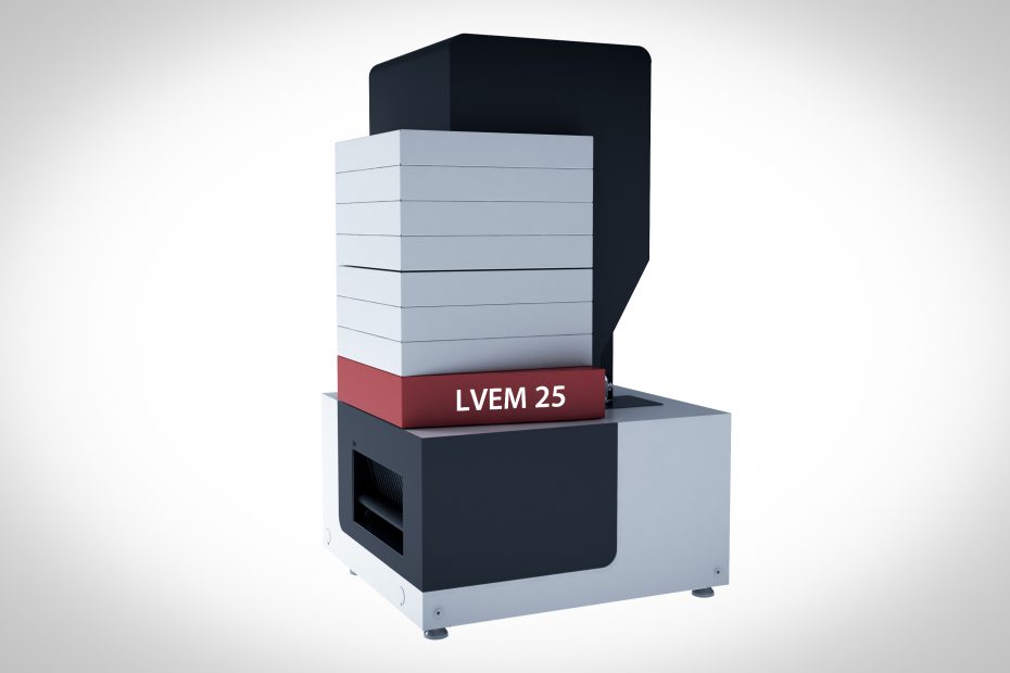
The low-voltage small transmission electron microscope mainly includes two models, LVEM5 and LVEM25.
Low-voltage small TEM LVEM5 and LVEM25 are electron microscopes with multiple functions integrated, with four different imaging modes – transmission electron microscope (TEM), scanning electron microscope (SEM), scanning transmission electron microscope (STEM), and electron diffraction ( ED), can provide virology researchers with multiple imaging modes required for characterization at the same time, and comprehensively analyze the structure and composition of virus samples.
Its main features are as follows:
LVEM5 (electron acceleration voltage: 5kV)
The LVME5 small low-voltage transmission electron microscope is a desktop transmission electron microscope. Its main features are as follows:
1. The volume is much smaller than the traditional large-scale transmission electron microscope, and the desktop design can be placed on any laboratory desktop;
2. Schottky field emission electron gun: high brightness, high contrast;
3. Multiple imaging modes: transmission electron microscope (TEM), electron diffraction (ED), scanning electron microscope (SEM), scanning transmission electron microscope (STEM) [1];
4. Resolution: 1.5nm (TEM); 10nm (SEM)
5. Observe biological samples without staining
LVEM25 (electron acceleration voltage: 10, 15, 25 kV)
1. Using a Schottky field emission electron gun, high brightness, and contrast
2. Multiple imaging modes: transmission mode (TEM), scanning transmission mode (STEM), electron diffraction mode (ED)
3. Observe biological samples without staining
LVEM uses a specially designed inverted Schottky field emission electron gun to provide high-brightness and high-coherence electron beams. The interaction between this low-energy electron beam and the sample is much stronger than the high-energy electrons in traditional transmission electron microscopes. This causes electrons to be strongly scattered by light organic materials, leading to an unusual differentiation of features.
In terms of virology research, the maximum magnification of the device is higher than the magnification of about 50,000 times that is usually required for observing viruses, and it still maintains a good resolution (<2 nm), which can meet the needs of virus morphology and structure research.
Compared with high voltage, the electron beam provided by the accelerating voltage of 5kV has a stronger interaction with the sample, has higher sensitivity to density and atomic number, and can still obtain a good sample image for a density difference as low as 0.005 g/cm3 Contrast, effectively improving the imaging quality of light element samples, suitable for virological research.
For the study of virus-like vectors, the low-voltage transmission electron microscope can directly observe the shape of virus-like vectors without negative staining, which helps researchers to quickly screen vectors and solve the problems of traditional electron microscope sample preparation and time constraints.
Advantages of Low-Voltage Compact TEM
- Small size and low environmental requirements, can be installed in any laboratory
The traditional large-scale transmission electron microscope is bulky, has strict requirements on the placement environment, is very sensitive to vibration, and needs to be installed on a good shock-absorbing foundation, so it can only be installed on the first floor or basement.
And due to the high energy consumption of large-scale transmission electron microscopes, many peripheral devices (such as water coolers, etc.) are required to maintain their operation, which usually occupy the entire laboratory.
The low-voltage small transmission electron microscope (LVEM) is different from the traditional electron microscope. It has no strict requirements on the placement environment. The size is 90% smaller than that of the traditional electron microscope. The power consumption is low. It does not need any external cooling equipment. It can be installed in any laboratory or Office desktop.
- High contrast, especially suitable for light element samples
Traditional TEM mostly uses electron beam acceleration voltage of 80-300kV. High-energy electron beams cannot distinguish similar densities and atomic numbers in light materials. It is difficult to obtain good contrast for light element samples (C, N, O samples, biological samples), which affects the image. quality.
The low-voltage small transmission electron microscope adopts a 5kV low-voltage design. The low-voltage electron beam has high sensitivity to density and atomic number, and can still obtain good image contrast for density differences as small as 0.005 g/cm3.
- Observation of biological samples without staining
When traditional electron microscopy observes biological samples, heavy metal staining is usually performed to provide contrast for the image. After the sample is stained with a heavy metal stain, the contrast is improved, but it also brings some problems:
1) In fact, all heavy metal dyes are harmful to our human body, and the operation must be very careful and strictly controlled;
2) The heavy metal stain interacts with the sample and distorts the true structure of the sample itself;
3) Heavy metal stains may completely destroy the sample due to their strong alkalinity.
Because of its low electron beam acceleration voltage, the small transmission electron microscope increases the interaction between the electron beam and the sample, thereby improving the image contrast. It can observe biological samples without heavy metal staining, avoid the false image caused by staining, and observe the real structure of the sample.
- Reduce the damage of the electron beam to the sample
Many samples that are sensitive to electron beams, such as carbon nanotubes, organic materials, biological samples, etc., are easily deformed or even decomposed under the traditional electron microscope. Therefore, it is impossible to carry out long-term detailed observation, or it is impossible to observe. The low electron beam acceleration voltage of the small transmission electron microscope reduces the damage of the electron beam to the sample, prolongs the observation time, and even sees samples that cannot be observed by the large electron microscope.
- Simple operation and low maintenance cost
Compared with the complex operation of the large-scale transmission electron microscope, the operation of the small-scale transmission electron microscope is much easier and does not require very professional operators. Even those who have never operated an electron microscope can easily get started with simple training. Its applicable units are not limited to scientific research institutes, and non-professional units such as enterprises and hospitals can also be equipped.
In use, no cooling water is required, no professional laboratory is required, and the maintenance cost is extremely low.
Disadvantages of low-voltage compact TEM
unable to achieve high resolution
In order to achieve angstrom-level high resolution (lattice, atomic-level resolution), the transmission electron microscope needs a high electron beam acceleration voltage and has strict requirements on the environment. Due to the low-voltage accelerating voltage of 5kV, the low-voltage small transmission electron microscope (LVEM) cannot achieve atomic-level resolution, and its resolution can only reach 1-2nm.
Summarize
Taken together, the results suggest that low-voltage microscopy is a suitable alternative to high-voltage transmission electron microscopy for virus diagnostic electron microscopy. In ultrathin sections, ultrastructural details of viral origin can be revealed. Imaging is fast enough to rapidly screen diagnostic samples with reasonable throughput. Taken together, the results suggest that low-voltage microscopy is a suitable alternative to high-voltage transmission electron microscopy for virus diagnostic electron microscopy.