Microscopes are ubiquitous instruments in every research laboratory. Whether pocket-sized or full-sized setups, microscopes are used in a variety of settings, including research, healthcare, education, manufacturing, and engineering.
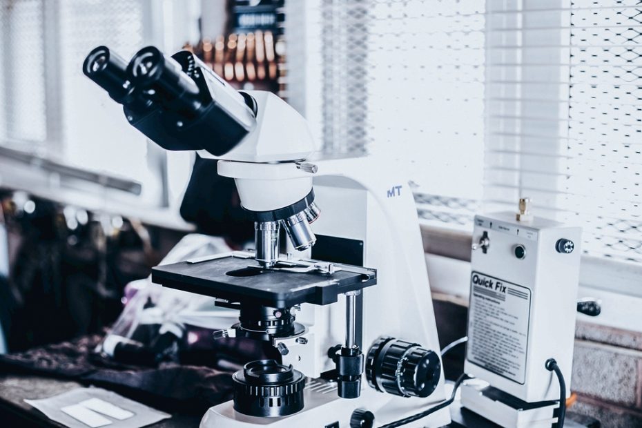
Both optical microscopes and digital microscopes are composed of objective lenses, eyepieces, a stage, spotlighting system, and a focusing mechanism.
The objective lens is located near the object to be observed to realize the magnification lens and the objective lens with different magnifications is installed on the objective lens converter to provide different magnification requirements.
A variation of the optical microscope is the digital microscope. These microscopes use a digital camera instead of eyepieces. The images are displayed in real-time on a computer screen. The invention of the USB port in the 1990s allowed for a wide variety of USB digital microscopes to suit every need.
The eyepiece is a lens located near the human eye to achieve magnification. Its magnification is relatively small, and the maximum magnification is only 20X. Generally, the size of the object under the microscope is determined by the objective lens.
The condenser is an important device to improve the influence of the light source on the imaging quality, and it is of great help to the resolution and image clarity of the microscope.
Optical microscope
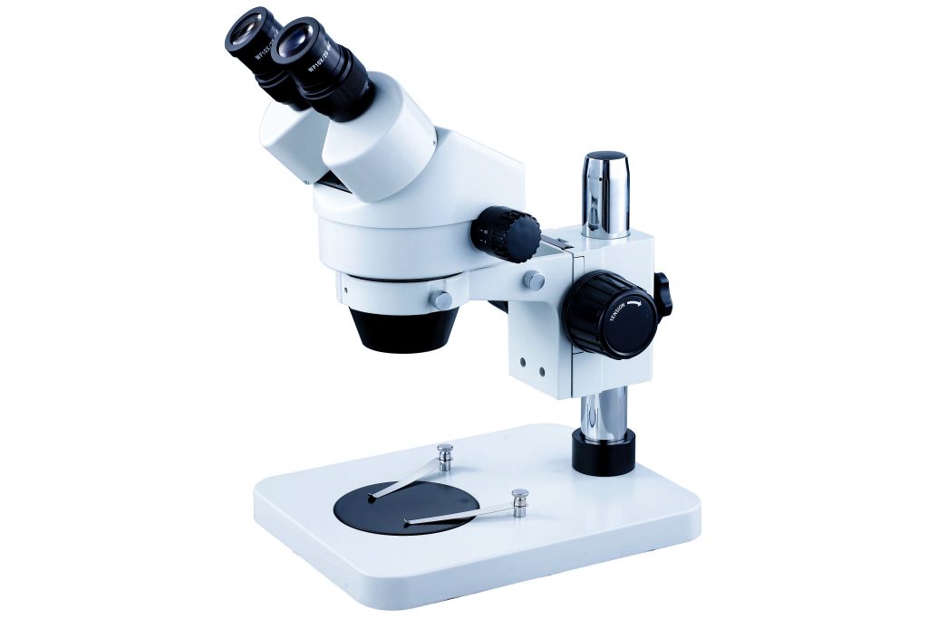
Optical microscopes use visible light and a lens system between the sample and the observer’s eye, which uses the human eye as a receiver to observe a magnified image.
The design and specifications of modern light microscopes can be very different from traditional simple microscopes that use sunlight as a light source. However, the basic principles are the same.
Principles of Optical Microscopy
A beam of light (from a light-emitting diode or light bulb) is focused on the sample to be studied. Light passes through it, through a set of objective lenses and eyepieces (eyepieces), and into the observer’s eyes.
Depending on the type of objective used, different magnifications are obtained. In today’s microscopes, the lenses are fixed in the stage condenser above and below the slide. When additional magnification is required, use objectives positioned above the sample.
Magnification depends on three variables
Three variables play a role in the magnification to be achieved. These are the angle of incidence, the angle at which light enters the lens, the curvature of the lens, and the index of refraction (RI).
Digital microscope
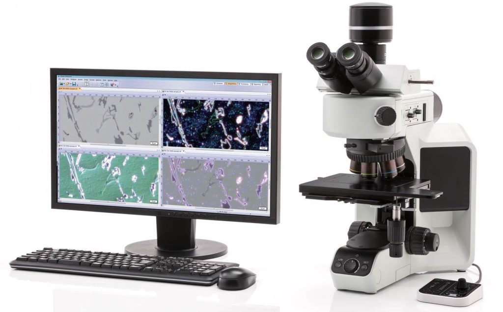
In simple terms, digital microscopes use a digital camera instead of eyepieces. The microscope is connected to a computer monitor and results are obtained in real time.
Principles of Digital Microscopy
Digital microscopes are often called video microscopes, which convert optical signals into electrical signals and image them on a computer, which greatly facilitates our observation and measurement of various experimental data of the object being inspected.
Digital microscopes can range in complexity from simple handheld versions to highly advanced systems. The latter uses complex software to perform advanced tasks.
Some of the advanced features available include image editing, 3D sample analysis, taking measurements, and report generation.
The difference between a traditional optical microscope and a digital microscope
Digital microscopes perform the same tasks as optical microscopes. However, there are other functions. Table 1 highlights the main differences between conventional optical microscopy (COM) and digital microscopy (DM). A key difference between a digital microscope and a conventional light microscope is that the former cannot directly view the sample through the eyepieces.
| Feature | Conventional optical microscopy | With higher magnifications, the depth of field becomes shallower |
| Depth-of-field | The depth of field in DM is 20-times more when compared to COM | COM lacks this capability. All specimens must be observed directly above |
| Variable-angle observation capability | COM lacks this capability. All specimens must be observed from directly above | DM achieves observation through 360 degrees. The sample need not be tilted |
| Three-dimensional imaging | Images using COM are two-dimensional. Surface characteristics are difficult to determine | DM gives three-dimensional and fully focused images |
| Quantitative data | COM does not provide measurements and other quantitative data | In DM, built-in measurement tools provide real-time measurements |
| Data Sharing | Not possible with COM | Images are easily recorded and stored on-site |
Why choose a digital microscope?
Digital microscopes mainly have the following advantages:
1. With the function of a microscopic camera, the observed microscopic effect is saved and formed into a graphic file, which can be circulated to relevant departments; the optical microscope can only be observed through the eyepiece, and cannot be used for microscopic photography.
2. Connected with various display devices such as computers/tablets/mobile phones/TVs/screens, multiple people can observe at the same time; optical microscopes can only be observed by one person.
3. The screen preview can reduce eye fatigue; optical microscopes need to observe through the eyepieces all the time, which is easy to cause excessive eye fatigue.
4. The imaging device of a digital microscope can perform functions such as measurement, the printing of graphic reports, and video recording; an optical microscope can only perform microscopic observation.
5. Digital microscope is a new era in the development of modern scientific instruments and meters and has many functions that optical microscopes do not have.
6. The digital microscope provides a large depth of field, allowing you to observe cell cultures precisely, even on uneven surfaces. Combine this with a deep compositing feature that allows the focus positions of a large number of images to be stacked, and you have high-resolution photos.
7. The digital microscope has a variable angle function and can perform 360° observation. You don’t have to tilt the sample. In addition, inspection times are greatly reduced.
8. Some digital microscopes have multiple illumination functions. Sometimes parts of the image may not be clear due to glare. By changing the direction of the ring light, glare is eliminated in the image. All this at the touch of a button.
9. You will be able to analyze your cell cultures in depth due to the magnification range offered by the digital microscope. Various magnifications are available from 0.1X to 6000X.
10. Digital microscopes can connect to cloud-based systems to facilitate remote work. This means you can use your computer to operate the device and observe your samples.
With remote monitoring, you can evaluate cell cultures inside the incubator. The key advantages here are: you can regularly evaluate your cells from a remote location; you can efficiently schedule visits to the lab; you store all relevant data and can analyze images from your desk.
11. Your digital microscope plays a vital role when you want to perform live imaging of cell cultures instead of using fixed cells.
In live-cell imaging, time-lapse microscopy is used to dynamically observe, track, and quantify processes in living systems. Such information is more likely to be of physiological relevance than information obtained from fixed cells.
In conclusion
Technology has come a long way since the invention of the microscope in the late 16th century. Light microscopy has traditionally been the workhorse in a variety of settings, including research. Digital microscopy has several unique features that give it advantages in a research setting.
Alternative Types of Microscopes
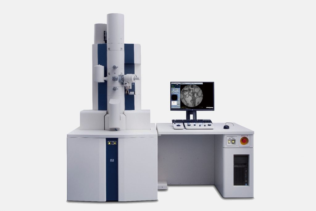
When the limitations of a light microscope are great, alternative types of microscopes may be more useful, and there are several other types of microscopes that can be used as light microscope alternatives. These include:
- Scanning Electron Microscope
- Transmission electron microscope
- Fluorescence microscope
- Atomic Force Microscope
- Scanning ion conductance microscope
- Scanning tunneling microscope
- UV microscope
- X-ray microscope
Unlike light microscopes, these types of microscopes do not use visible light to view samples.