
The lenses of an electron microscope correspond to a converging lens, an objective lens, an intermediate lens, an eyepiece, and a projector lens. Compared with optical microscopes, even with the same name “lens”, the structure of the lens itself is different because electron beams are used as the light source.
To focus the electron beams, instead of using materials such as glass, we use electromagnetic action such as magnets. Therefore, “electron lenses” such as “electrostatic lenses” and “electromagnetic lenses” are used. There are many types of electronic lenses, but what they have in common is the use of electromagnetic effects.


Figure: Comparison of optical lens and electronic lens
Scanning Electron Microscopy (SEM) uses electron beams to image samples with nanoscale resolution. The filament emits electrons, forming parallel electron beams. The electron beam is then focused on the sample surface through a lens. How do electronic lenses work? What types of electronic lenses exist? How does an electron lens focus electrons?
Scanning Electron Microscopy: Electron, Electron Beam, and Electron Lens
Electrons in a scanning electron microscope (SEM) are released from the filament and then parallel to the electron lens.
The electron beam travels through the column—consisting of a set of lenses that focus the electron beam onto the sample surface.
Electron microscope lenses can be electrostatic or magnetic, depending on whether they use electrostatic or magnetic fields to focus the electron beam. To better understand how these lenses work, let’s step back and look at how electrons are deflected by an electrostatic field.
Guide plate
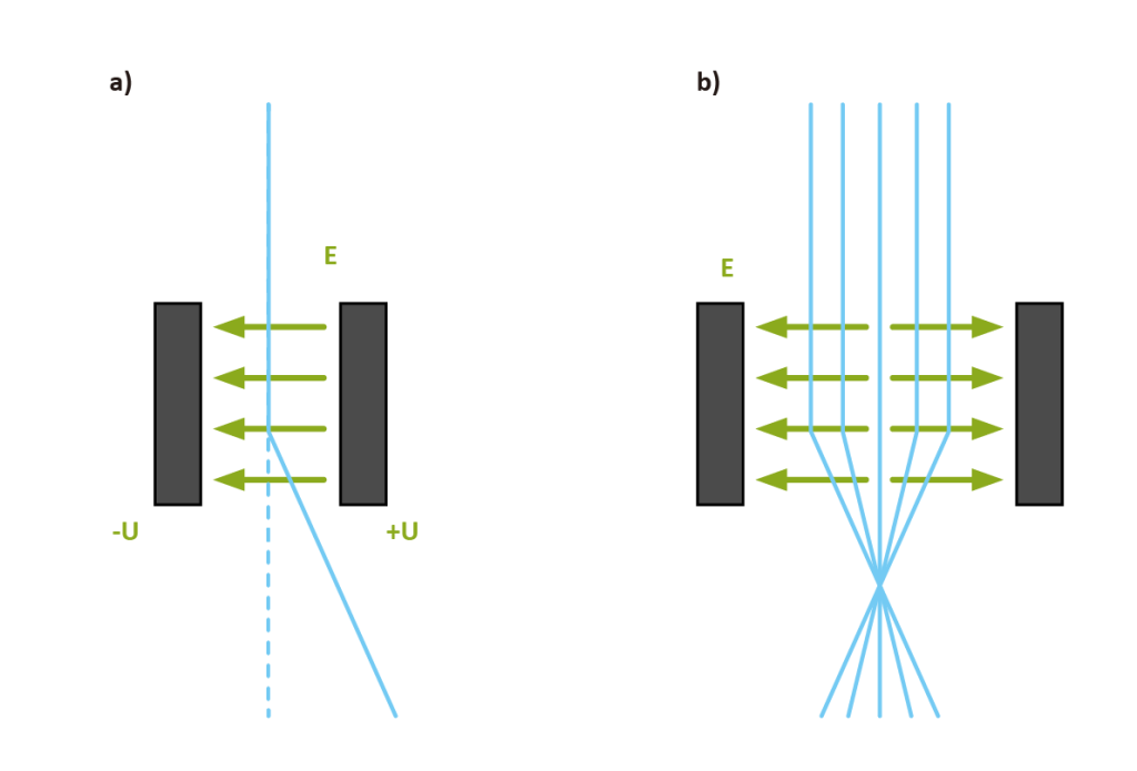
Electrons are negatively charged particles that travel through the high-energy column. One way to deflect these particles is to pass them through an electric field generated by two plates at potentials +U and -U, as shown in Figure 1a.
Under the influence of the electric field, the deflection angle of the electron depends on the electron energy, the electric field between the plates, and the length of the plates.
The faster or more energetic the electron, the smaller the angle of deflection. The higher the electric field, the longer the plate and the greater the deflection angle. A device consisting of two plates of different potentials is called a guide plate.
In order to obtain an electrostatic lens, reflective guide plates can be considered, so that electrons moving on the optical axis can be focused on the same point, as shown in Figure 1b.
The electric field only exists when the electrons start to enter and end the electron journey, how can we get the lensing effect as shown in Figure 1b? The answer to this question is that as long as there is a lensing effect, the energy of the electron beam will change, which means Electrons are either accelerated or decelerated. This can be done with different potentials around the electron beam.
Electrostatic lens
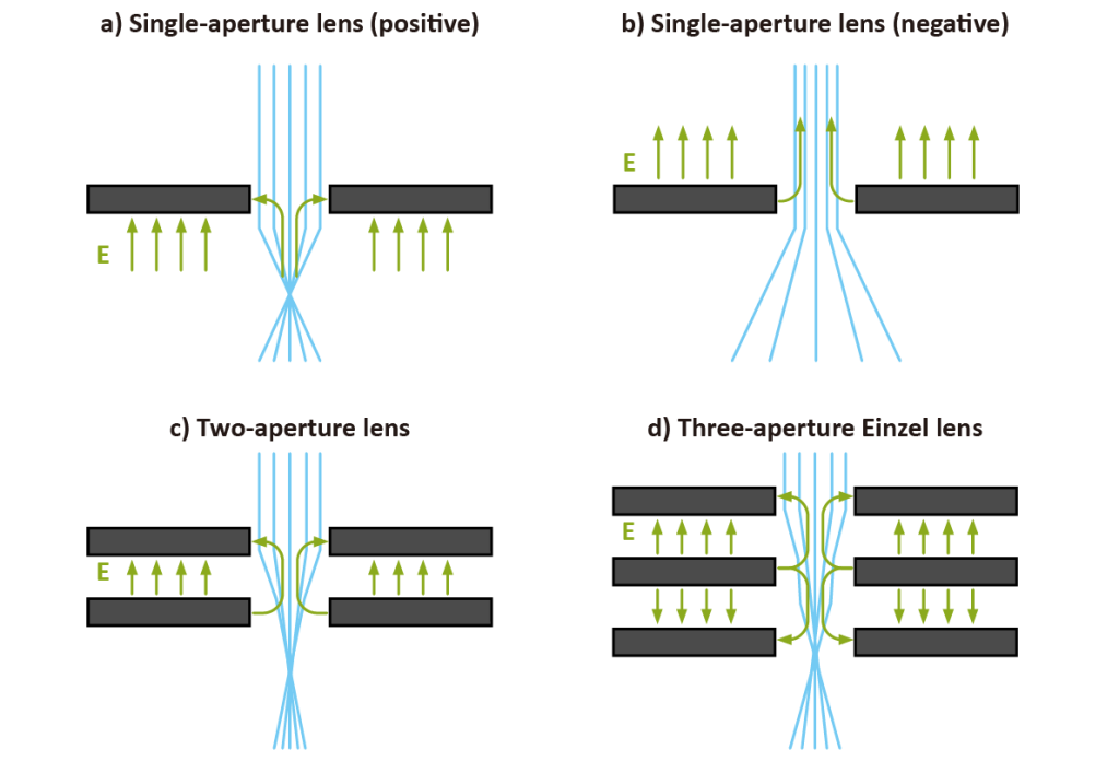
An electrostatic lens consists of a metal plate connected to a high voltage through which electrons pass through the electron lens. Single aperture lenses consist of a single metal plate under high voltage.
The single-hole lens can not only terminate the accelerating field but also generate the accelerating field. In this example, the lens is positive, which means that the electron beams converge, as shown in Figure 2a, while in the second case, the lens is negative, which means that the electron beams diverge, as shown in Figure 2b.
A double-aperture lens consists of two metal plates, aligned at the apertures, with different electrical potentials. Figure 2c shows an accelerated two-hole lens with the electric field between the two plates on the top plate.
Electrons entering this lens feel a strong magnetic field that draws them closer to the optical axis. As they pass through the second plate, the electrons feel an opposing force pushing them toward the lens. In general, this is a positive lens, and the electron beam is focused on the plane below the second plate.
A triple-aperture Einzel lens consists of three plates with aligned apertures, which can have the same or different diameters. In electron optics, Einzel lenses are often used to have equal electron potentials at the entrance and exit of the lens.
In Figure 2d, an accelerated Einzel lens is shown, with three electrodes producing three lenses: the first and third are positive and the second is negative. The overall lens is positive and the beam is focused on the plane below the third lens.
Electromagnetic lens
Electromagnetic lenses use the Lorentz force, which is proportional to the charge and velocity of electrons, to deflect them. A magnetic lens consists of a metallic body (called a ferromagnetic circuit) that ends with two pole pieces.
The magnetic field is generated by a coil placed on top of the ferromagnetic circuit, as shown in Figure 3. The strength of the lens can be changed by changing the magnetic field B, which is done by changing the geometry of the rods, the distance between the rods, and the coil current.

Scanning Electron Microscope Electron Lens
Electron beams consist of electrons released by an electron source with a set of lenses. The electrons are compressed into a beam by the electron lens, and then focused on the sample surface by the final lens, which is also called the objective lens, as shown in Figure 4. The electron source is tilted, and the scanning electron beam over the sample is produced by the coil of the electron source and the last lens on the right.
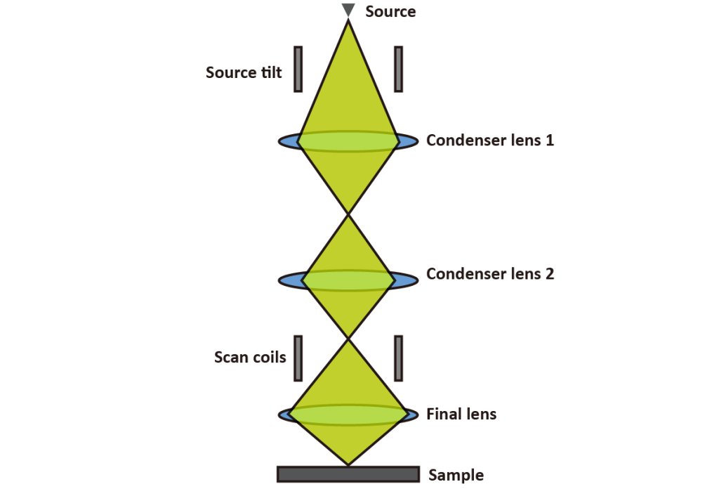
Figure 4: Electron column
All scanning electron microscopes (SEMs) — whether we’re talking benchtop or floor standing — have a column with an electrostatic lens and an electromagnetic lens.