Introduction to transmission electron microscopy
Transmission electron microscopy (TEM) is a technique in which a beam of electrons is passed through a sample to form an image. The sample is usually an ultrathin section less than 100 nanometers thick or a suspension on a grid. As the electron beam passes through the sample, the interaction of the electrons with the sample forms an image. The image is then magnified and focused onto an imaging device, such as a fluorescent screen, photographic film, or sensor. The sensor may be a fluorescent material attached to a charge-coupled device.
Because electrons have smaller de Broglie wavelengths, transmission electron microscopy can image at much higher resolution than optical microscopy. This allows the instrument to capture minute details – even as small as a column of atoms, which is thousands of times smaller than objects that can be resolved in an optical microscope. Transmission electron microscopy is a major analytical method in the physical, chemical and biological sciences. TEMs are used in cancer research, virology, materials science, and pollution, nanotechnology, and semiconductor research.
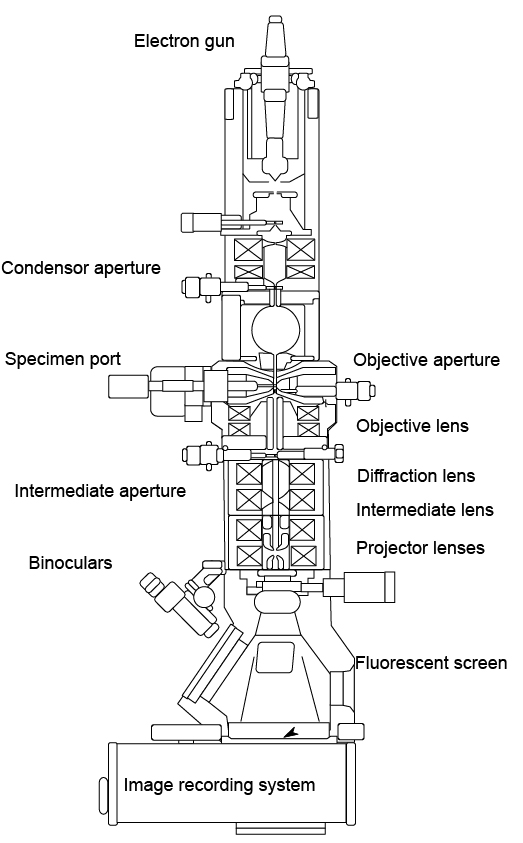
Structure of a transmission electron microscope
TEM mainly consists of five parts: lighting system, imaging and amplification system, observation and recording system, vacuum system, and adjustment system:
1. Lighting system
Mainly includes electron gun and condenser
Electron gun: emits electrons and consists of a cathode, a grid, and an anode. The electrons emitted by the cathode tube form a ray beam through the small hole on the grid, and are accelerated by the anode voltage and then directed to the condenser, which accelerates and pressurizes the electron beam; the condenser: gathers the electron beam and can be used to control the illumination intensity and Aperture angle.
2. Imaging and magnification systems
It mainly includes a sample room, objective lens, intermediate mirror, and projection mirror. The sample room is used to place the sample to be observed and is equipped with a tilting table to change the angle of the sample. It is also equipped with heating, cooling and other equipment; the objective lens: is for very high magnification. A tall, short-distance lens that magnifies electron images. The objective lens is the key to determining the resolution and imaging quality of a transmission electron microscope; the intermediate mirror: a weak lens with variable magnification, which is used to amplify the electron image twice. By adjusting the current of the intermediate mirror, the image of the object or the electron diffraction pattern can be selected for magnification; the projection mirror: a high-magnification strong lens used to amplify the intermediate image and then image it on the fluorescent screen.
3. Observation and recording system
Mainly includes a binocular microscope, observation room, fluorescent screen, and photography room.
4. Vacuum system
It mainly includes a vacuum pump and a vacuum column. The vacuum column appears as a sealed cylindrical container, and the vacuum pump is used to generate a vacuum within the vacuum column.
5. Tuning system
It mainly includes condenser diaphragm, objective lens diaphragm, intermediate mirror diaphragm, etc.
How transmission electron microscopy works
The electron beam emitted by the electron gun passes through the condenser along the optical axis of the mirror body in the vacuum channel, and is condensed into a sharp, bright, and uniform light spot through the condenser, which illuminates the sample in the sample chamber; after passing through the sample The electron beam carries the structural information inside the sample. The amount of electrons transmitted through the dense areas of the sample is small, and the amount of electrons transmitted through the sparse areas is large. After the focus adjustment and primary magnification of the objective lens, the electron beam enters the lower intermediate lens and The first and second projection mirrors perform comprehensive magnification and imaging, and the finally amplified electronic image is projected on the fluorescent screen in the observation room; the fluorescent screen converts the electronic image into a visible light image for the user to observe.
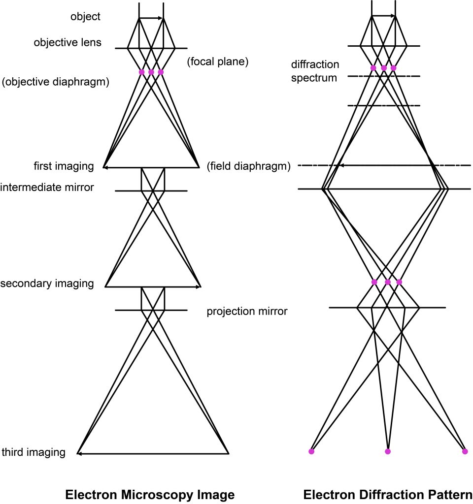
The TEM imaging process conforms to the Abbe imaging principle
- When a parallel electron beam is an incident on a periodic structure sample, diffraction occurs.
- After being focused by the objective lens, a diffraction maximum is formed on the back focal plane.
- The secondary waves emitted by each diffraction maximum are coherently imaged on the image plane.
- The image on the image plane passes through the intermediate lens group, and the projection lens group is magnified twice and projected onto the fluorescent screen, which is called the third-level magnification of the object.
- By changing the current of the intermediate mirror, that is, changing the focal length of the intermediate mirror, so that the object plane of the intermediate mirror moves to the back focal plane of the objective lens, the process of image transformation into a diffraction spectrum can be seen on the fluorescent screen.
Features of transmission electron microscope
Due to limitations of sample preparation technology, for most biological samples, only a resolution of 2 nm can generally be achieved.
The resolving power of transmission electron microscope images depends not only on the resolution of the electron microscope itself but also on the contrast of the sample structure.
The light source used in transmission electron microscopy is electron waves. The wavelength has no color reaction in the non-visible light range. The image formed is a black-and-white image, requiring the image to have a certain contrast.
Biological tissues and cell components mainly comprise light elements such as C\H\O\N. Their atomic numbers are low, their electron scattering ability is weak, and the differences between them are very small. The image contrast under the electron microscope is generally low.
Due to the weak penetration of electron beams, samples must be sliced into ultrathin sections. The observation surface is small, the net carrying capacity is 3mm, and the ultra-thin sectioning range is 0.3-0.8mm.
Strong irradiation of electron beams can easily damage the sample, causing deformation, sublimation, etc., or even breakdown and rupture, which may cause artifacts in the observed structure.
The transmission electron microscope barrel must be kept in a vacuum during observation. In order to ensure that the sample is not damaged under vacuum, the sample must be free of moisture. Therefore, living biological samples cannot be observed.
Biological sample preparation is complex. During the multi-step sample preparation process, samples are prone to structural changes such as shrinkage, expansion, fragmentation, and loss of content. 4
Six main functions of transmission electron microscopes
- Use mass-thickness contrast (also called absorption contrast) images to observe the general morphology of the sample
- Use techniques such as electron diffraction, micro-area electron diffraction, and convergent beam electron diffraction to conduct phase analysis on samples to determine the phase, crystal system, and even space group of the material.
- High-resolution electron microscopy can be used to directly “see” the structural projection of atoms or atomic groups in a specific direction in the crystal, and determine the crystal structure.
- Use diffraction contrast imaging and high-resolution electron microscopy technology to observe structural defects in crystals, determine the types of defects, and estimate defect density.
- Use the energy-dispersive X-ray spectrometer or electron energy loss spectrometer attached to the transmission electron microscope to analyze the micro-area chemical composition of the sample.
- Use a transmission electron microscope with a scanning accessory and an energy-dispersive X-ray spectrometer, or use a transmission electron microscope with an image filter, to analyze the elemental distribution in the sample to determine whether there is component segregation in the sample
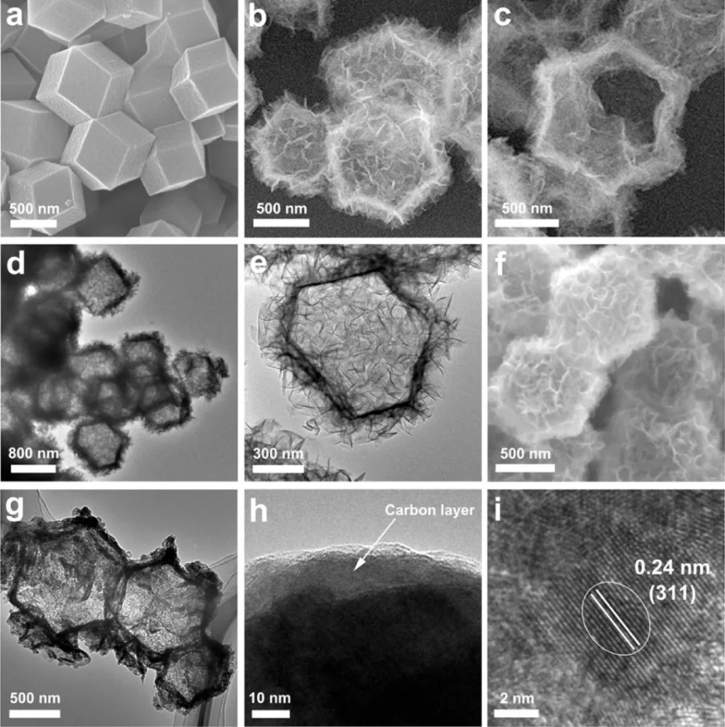
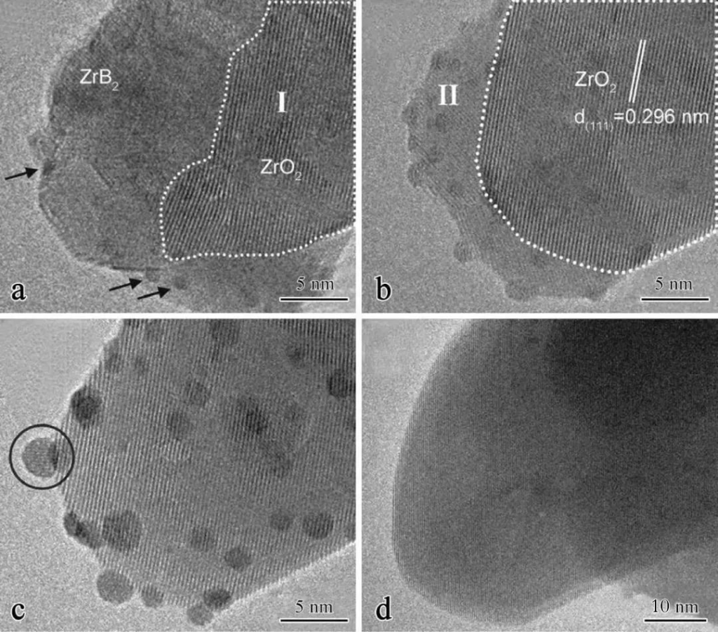
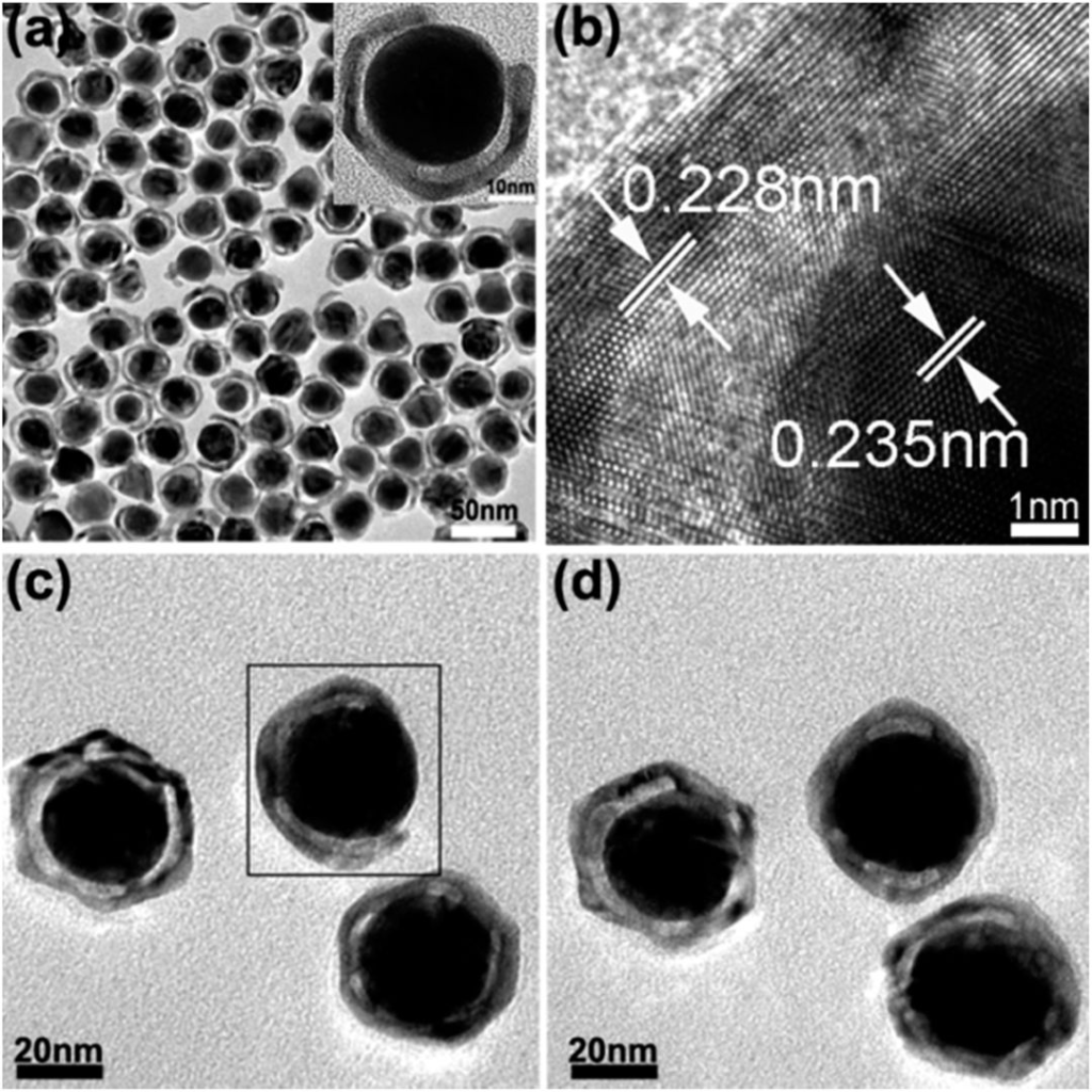
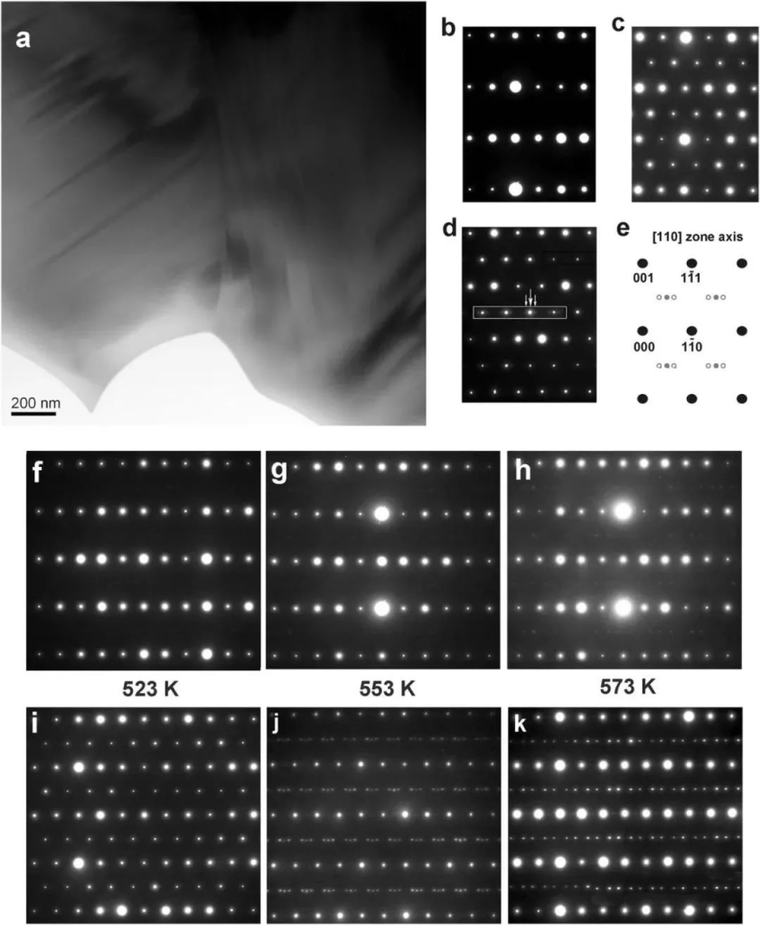
Appreciation of typical morphology of transmission electron microscope