What is differential interference microscopy?
A differential interference microscope (DIC) is also called the Nomarski contrast microscope. Its advantage is that it can display three-dimensional projection images of structures. Compared with phase contrast microscopy, the specimen can be slightly thicker and the refractive index difference is larger, so the image has a stronger three-dimensional effect. Differential interference microscopy uses plane-polarized light as the light source. The light is divided into two beams after being refracted by the prism, passing through adjacent parts of the sample at different times, and then passing through another prism to merge the two beams of light, thereby converting the small difference in thickness in the sample into a difference in light and dark.
Four major characteristics of differential interference microscopy
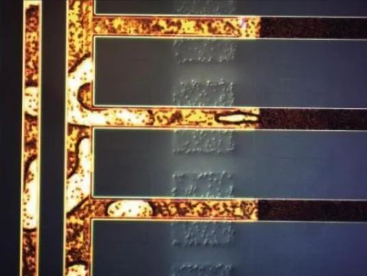
- 1. The preparation requirements for metallographic samples are reduced. For some samples, it can even be observed by polishing without corrosion treatment. The advantage is that the true state of the sample surface can be observed, such as polishing the sample under vacuum. Martensite phase transformation, the embossment of martensite phase transformation can be observed without corrosion.
- 2. The observed surface has obvious concavity and relief, and the relative hierarchical relationship between the various components of the sample can be displayed. Correct judgments can be made on particles, cracks, holes protrusions, etc., which improves the quality of metallurgy. It improves the accuracy of phase inspection and also increases the contrast between phases.
- 3. When you use the differential interference contrast method to observe samples, you will see many details that cannot be seen in bright fields. Some structural details or defects that are difficult to distinguish in bright field can be easily judged by contrast enhancement through differential interference.
- 4. The differential interference contrast method is based on the traditional cross-polarization method, and cleverly uses the improved DIC prism and complementary color filter (^-plate) based on the Wollaston prism to make the observed sample use optical interference. This method produces rich microphotography. Because the effect of differential interference contrast, the relief of sample details, and the color can be adjusted, it is superior to cross-polarization.
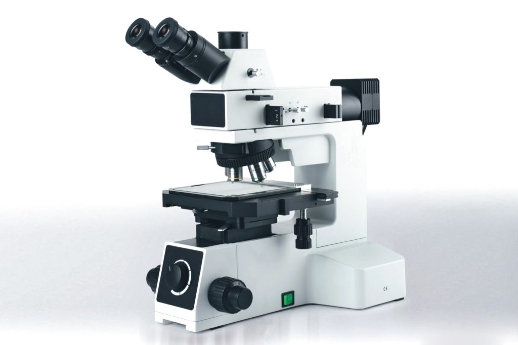
Four product observation methods of differential interference microscopy
1. DIC differential interference observation:
Based on orthogonal polarization, DIC differential interference observation can be performed by inserting a prism. Using DIC technology, the slight height difference on the sample surface can produce an obvious relief effect, greatly improving the contrast of the image. It can be matched with the DIC function The objective lens makes the interference color consistent across the entire field of view, the differential interference effect is excellent, and the DIC effect of the high-magnification objective lens is also good.


2. Bright field observation (reflection):
The telecentric Kolar reflection illumination system, coupled with the newly designed infinity long working distance plan achromatic metallographic objective lens, can obtain clear, flat and bright high-quality microscopic images from low to high magnification.
3. Darkfield observation:
Pull the dark field lighting lever to the designated position to use the dark field function, which allows you to observe various scratches, impurities and other minor defects on the surface of the object. The built-in attenuator reduces the irritation to the eyes caused by the dramatic changes in light when switching between light and dark fields. .
4. Simple polarized observation:
Insert the polarizer and analyzer insert the plate into the designated position of the illuminator to perform simple polarization observation. The analyzer can be divided into fixed type and 360° rotating type.

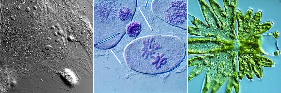
The differential interference microscope can not only observe colorless and transparent objects, but also the image presents a relief-like three-dimensional effect and has certain advantages that phase contrast microscopy cannot achieve, and the observation effect is more realistic.
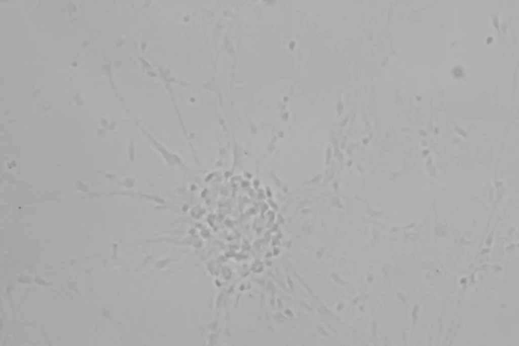
Differential interference contrast is an excellent solution for brightfield illumination imaging of unstained transparent biological samples.
Differential interference contrast (DIC) is an excellent solution for brightfield illumination when imaging unstained, transparent biological samples using a microscope. Brightfield images of samples often lack structure and detail because there is little variation in the intensity of light absorption in localized areas. Staining does cause a difference in the intensity of light as it passes through the sample, but staining is often toxic. DIC microscopy utilizes gradient changes in optical path length and phase shift to reveal transparent structures.
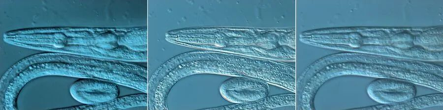
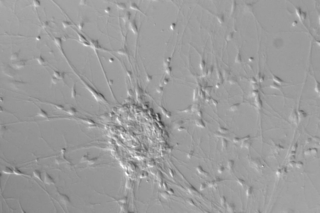
1. Relief-like image under polarized light
In brightfield microscopy, unstained samples are often inconspicuous and lack detail. In reality, they simply interact with the incident light and undergo a phase shift that is invisible to the naked eye. Staining causes amplitude shifts and differences in passing light intensity, but more importantly, this method only works on dead material. Specimens or structures that cause changes in the amplitude of light waves are called complex-amplitude objects.
Differential interference contrast (DIC) microscopy uses gradient changes in optical path length and phase shift to allow researchers to see phase objects when looking with a microscope. A sample or its structure that causes a phase shift when light passes through it is called a phase object. In this way, living cells and organisms can be observed with sufficient contrast and resolution. DIC can produce images with a relief effect, where the structure and features of the specimen appear to cast shadows. Because of optical sectioning, these relatively thick samples are imaged without halo artifacts.
For DIC microscopy, polarized light is used only to illuminate the sample. Polarized light is dispersed into two different rays located in orthogonal polarization planes. The two rays are very close to each other. Different phase shifts occur when they refract or scatter differently within the sample. If these rays of light rejoin, they interfere with each other. The light is now elliptically polarized. This polarized light becomes an amplitude shift when passed through the analyzer. In this way, the wavelength can be phase-shifted by 1/200 of the entire wavelength (or even 1/1000 when using a camera), so that the entire wavelength can be seen.
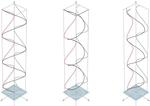
2. Oscillation of light waves
For unpolarized light, light waves oscillate in all directions relative to the axis of propagation. However, for linearly polarized light, all light wave oscillations have the same polarization angle or plane. Circular polarization is another possible type of polarization. Light wave oscillations follow circular motion and have a stable absolute value, while the direction changes at a constant angular velocity. Elliptically polarized light is a mixture of linearly polarized light and circularly polarized light.
Polarized light can be produced by materials or devices called polarizers. If a similar component is used to determine the plane of vibration, it is called an analyzer. There are absorption polarizers and spectroscopic polarizers.
3. Light path in DIC microscope
In a DIC microscope, polarized light is generated through a polarizer located in front of the condenser. This polarized light is divided into two linearly polarized lights perpendicular to the polarization plane through a DIC prism, and this DIC prism is also called a Wollaston prism. A Wollaston prism can be located in the plane of the condenser. A standard Wollaston prism is made from two wedges of birefringent material, both bonded to a base and having optical axes parallel to the outer surface and perpendicular to each other.
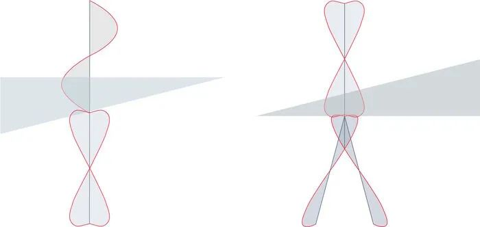
Polarized light passing through this prism is sheared into two closely spaced light waves with different deflection angles: a normal wave and an abnormal wave. They have perpendicular planes of polarization. For these two waves, the medium behaves as if it has two The refractive index is the same. Wavefront shearing occurs between two prisms at the interference plane located at the intersection of their refractive indices. The light waves are spatially separated by the shear angle. For all adapted light waves on the entire prism, their directions and spacing are the same. The space between the two light waves must be smaller than the resolution limit of the microscope.
As a result, the sample is illuminated by closely spaced light waves that enter as lateral displacements. If these two light waves interact with parts of the sample that have different refractive indexes or thicknesses, their optical path lengths will be different when they are transmitted out of the sample. The optical path length is the product of the refractive index and the thickness between two points on the optical path. Therefore, it is related to the transmission time and speed of light. Since the optical path length is related to the transit time of the light, a phase shift occurs between the two matched light waves.
After the light waves pass through the sample, they pass through another Wollaston prism, which brings them back together. At this point, they interfere with each other to form elliptically polarized light, which then passes through the analyzer. Only light waves with a distinct plane of polarization can pass through, so different light waves produce different amplitudes. The phase shift translates into an amplitude shift, resulting in different light intensities in the acquired images.
For modern DIC microscopy, Wollaston prisms are frequently modified. One example is the Nomarski prism. It also consists of two birefringent wedges, but only one wedge is identical to the wedge of the Wollaston prism. The other wedge is modified so that the optical axis is in a tilted position. This difference causes the interference plane to lie outside the prism. Therefore, this prism can be located outside the objective aperture plane, making DIC microscopes more convenient to use.
For DIC microscopy, differences in phase shifts between adjacent object points contribute to imaging. The details of the sample, if there are gradient changes in refractive index or thickness, can achieve a clearer visual representation. Because the beams are polarized perpendicular to each other, differences in the image can be seen by rotating the microscope stage.
It is important to remember that when performing DIC microscopy, never use samples together in plastic vessels, as polymers such as plastic have a depolarizing effect on light and can distort contrast.
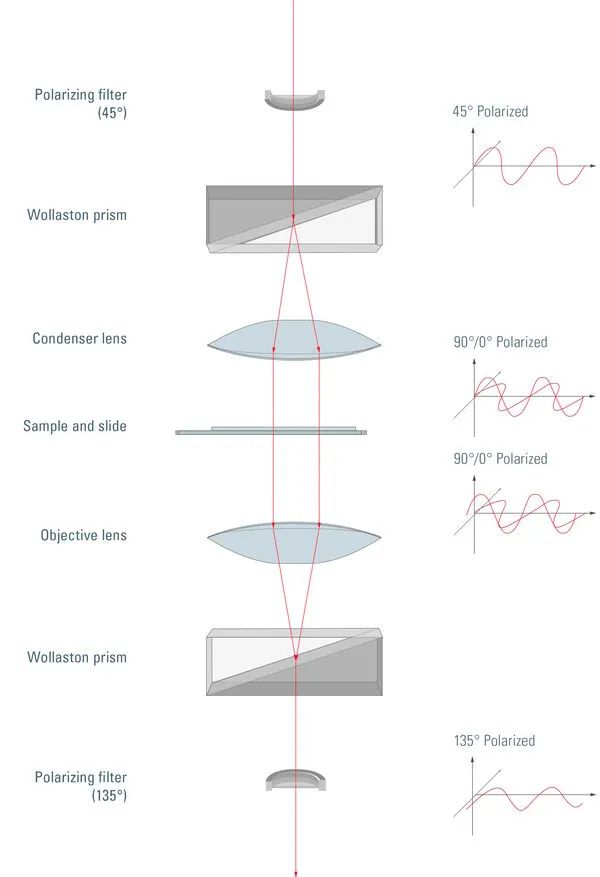
Unpolarized light passes through a polarizing filter and is polarized at 45°. After passing through the Wollaston prism, the light will be divided into vertical polarization components, one of which is 0° polarized and the other is 90° polarized. A condenser then directs the light through the sample. The sample is illuminated by two coherent parallel light rays with different polarization angles. This essentially produces two slightly offset brightfield images of the sample, one with 0° polarized light and the other with 90° polarized light. Due to the different polarization angles of the light, these images do not produce any constructive or destructive interference. Each ray traverses a different optical path length because there may be thickness or refractive index differences where it passes through the sample. The end result is a phase shift of one ray relative to the other. After passing through the objective lens, the vertically polarized light recombines into a single polarized light at the 135° position. Depending on the difference in optical path length, the interference of the two rays now produces a brightening or darkening of the sample area, making the structure appear almost invisible under brightfield illumination. Finally, a 135° polarizing filter (also called an analyzer) filters out the directly transmitted light without any phase shift.