Overview
1. An upright microscope can be used for most samples, but large-volume samples or samples that need to be placed from above during imaging require an inverted microscope.
2.The reflected illumination mode is suitable for opaque or thick samples, while the transmitted illumination mode is suitable for transparent or thin samples.
3. The bright field mode is most suitable for observing the absorption characteristics on the sample, while the dark field mode is suitable for observing the scattering characteristics on the sample.
The widespread use of optical microscopes goes far beyond the limits of what people can see with the naked eye. It is precisely because the use of optical microscope technology promotes the development of technology, so what is an optical microscope? What lighting technologies are available for optical microscopes? In this article, the editor will introduce optical microscopes and the lighting technology of optical microscopes.
01 Introduction to optical microscopy

In this article, we will introduce some of the most common light microscopy terms, including upright and inverted microscopes, reflected and transmitted illumination modes, and brightfield and darkfield illumination modes, and discuss their applicability to different research areas.
02 Upright microscope and inverted microscope
An important consideration when choosing a microscope is the position of the objective relative to the sample. In an upright microscope, the objective lens is located above the sample; in an inverted microscope, the objective lens is located below the sample. Despite differences in structure and appearance, they have the same imaging capabilities for samples and can be applied to different sample types.
When observing a sample with an upright microscope, the sample surface is facing upward (toward the objective lens located above the sample stage). Most samples can be imaged with an upright microscope that can accommodate slides, well plates, and specialized temperature, pressure, or electrochemical sample stages. However, there are two situations where an upright microscope is not suitable, namely when the sample is too large to be placed under the objective lens or when the sample needs to be placed from above during imaging. In both cases, an inverted microscope is required.
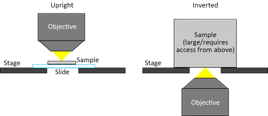
In an inverted microscope, the sample surface faces downwards (toward the objective lens located below the sample stage), which means that at least this side of the sample must be flat. An example of where inverted microscopes are better suited for live cell imaging is if the sample is a live cell culture system (where the cells sink to the bottom of the culture system and require access from above to exchange liquid media).
03 Reflected lighting and transmitted lighting
Because a range of samples with varying optical properties, from opaque semiconductor chips to translucent tissue cultures, can be viewed with the microscope, most users can choose either reflected or transmitted illumination mode depending on their specific sample. As shown in Figure 2, this is a schematic diagram of the light path in the two illumination modes of an upright microscope. In addition to this, inverted microscopes also have these two illumination modes.
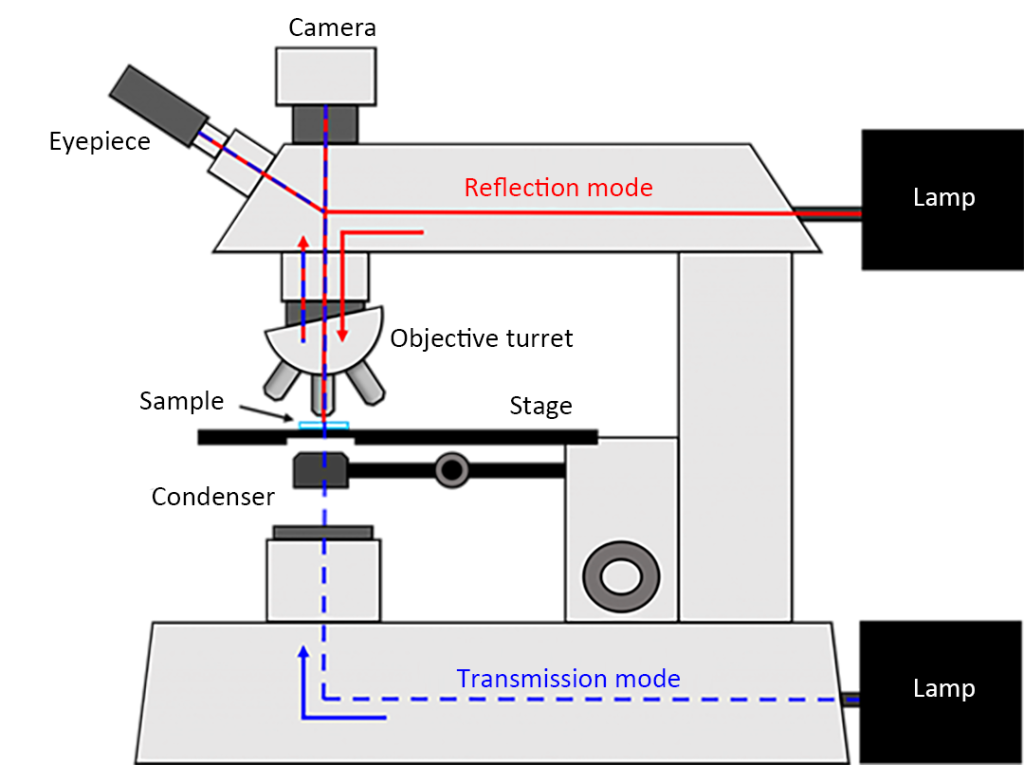
As shown by the red light path in Figure 2, the reflected illumination mode is considered the best mode for visualization of any opaque sample such as semiconductor chips, polymers, paints, paper, metals, and pharmaceuticals. When observing a sample using reflected illumination mode, light passes through the objective lens to the sample surface and then re-enters the objective lens after being reflected from the sample. Upon re-entering the objective lens, the light reaches the camera at the same time, enabling visualization of the sample.
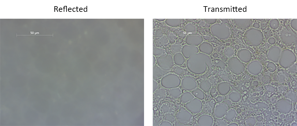
When imaging very thin or transparent samples, such as cells or tissue cultures mounted on glass slides, transmitted illumination is preferred. As shown in the blue light path in Figure 2, when using transmitted illumination, light passes through the sample and enters the objective lens, allowing for better observation of the morphology of the transparent sample. In transmission mode, since the incident light does not pass through the objective as in reflection mode, the light from the transmission is collected by a condenser located on the opposite side of the objective and focused onto the sample. In microscopes with two illumination modes, the direction of the light path can be changed by pressing a toggle switch on the frame. Figure 3 shows the samples under two lighting modes, in which the contrast of transmitted illumination is better than that of reflected illumination.
04 Brightfield lighting and darkfield lighting
There are two main modes of illumination in light microscopy, brightfield and darkfield. In the reflected brightfield illumination mode shown in Figure 4, light is reflected from the semi-silvered mirror to the center of the objective lens. In the reflected dark field illumination mode, the light baffle is connected and the reflector becomes a fully silver-plated annular reflector, allowing the light beam to move downward along the edge of the objective lens.
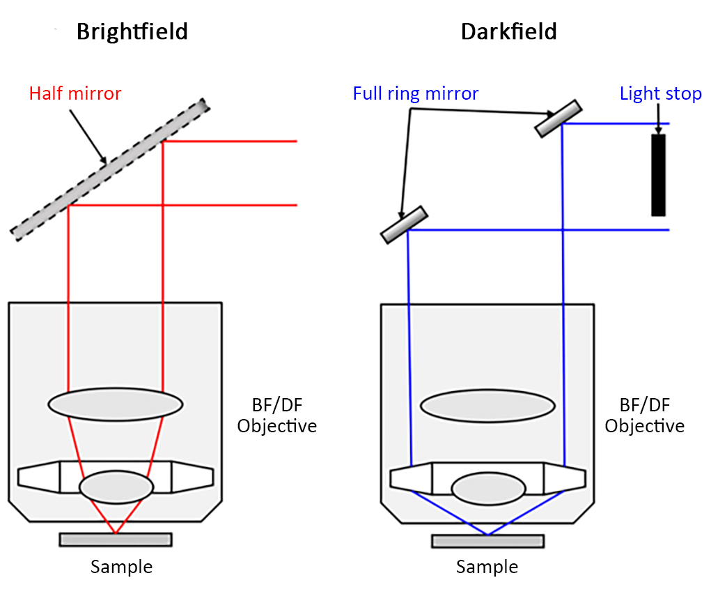
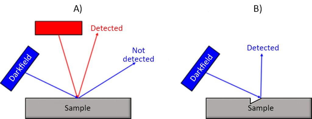
The two lighting modes differ primarily in the angle of incident light. Generally speaking, the bright field is illuminated at an acute angle between 45 degrees and 90 degrees from the sample horizontal plane, while the dark field is illuminated at an oblique angle between 0 degrees and 45 degrees, as shown in Figure 5. In brightfield mode, acute-angle light is mostly reflected back to the objective, if not absorbed. This means that absorbing features will appear dark against a bright background. In dark field mode, due to the oblique angle of incidence, light is usually not reflected into the objective lens, resulting in a dark background. Darkfield mode is used to detect defects and edges in the sample that cause incident light illuminated at an oblique angle to be scattered into the objective.
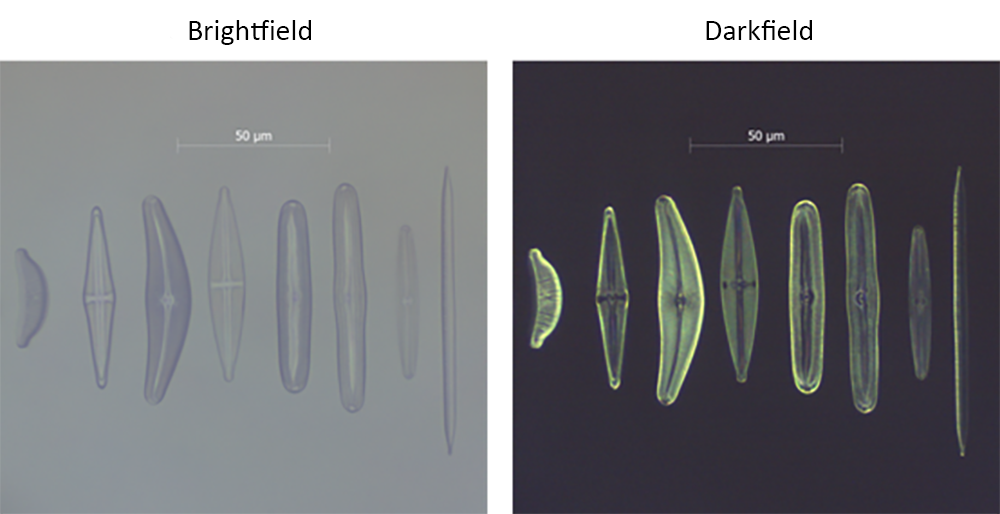
Darkfield requires specialized optics, and for reflective darkfield (as shown in Figure 4), specialized darkfield objectives and nosepieces are required. Transmitted darkfield is also available, requiring a darkfield condenser. As shown in Figure 6, the sample consists of various diatoms fixed on a glass slide, where darkfield illumination provides better contrast than brightfield illumination. As can be seen from the darkfield illumination images, the edges and fine structures of the diatoms that scatter light into the objective contrast well with the background, and the contrast is significantly improved relative to the brightfield mode.
05 Conclusion
Upright and inverted microscopes, reflected and transmitted illumination modes and brightfield and darkfield illumination modes are the most common terms used in optical microscopy. Before performing spectral analysis, different illumination modes can be selected for different types of samples. This not only ensures visualization of the sample, but also allows the user to find features of interest on the sample before imaging.