The lighting method in a microscope is an important factor affecting the quality of observation, photography, and measurement results. The correct lighting method cannot reduce the brightness and resolution, and there must be no spots or unevenness during lighting. The specific lighting method, also called the microscopic examination method, will be described later.
1. Microscope lighting method:
Specimen illumination methods are critical for microscopy. Illumination methods are broadly divided into transmitted illumination and incident illumination. An appropriate lighting method must be selected based on the specimen and purpose.
2. Transillumination
Illuminating the back of the specimen. This method is suitable for biological observation of colorless transparent cells and other samples and is generally used in biological microscopes. There are two types of transmitted illumination: brightfield and darkfield:
– Bright-field lighting
Typically, this method works by illuminating the back of the sample to make it transparent and observe it. Against a bright background, this method can display parts that are darker than the background.
– Darkfield lighting
Like brightfield illumination, this method illuminates the backside but blocks the transmission of direct light, revealing the outline of the specimen against a dark background. This method is suitable for observing low-contrast cells and must be used with a darkfield condenser.
3. Incident lighting
Illuminating the front of the sample. The method is suitable for observing 3D objects such as materials and other industrial samples, as well as opaque samples. This type of illumination method is generally used in stereomicroscopes.
4. The lighting system of the microscope should meet the following basic requirements:
- The light source has sufficient lighting brightness.
- Ensure strong and uniform illumination within the entire field of view being observed on the specimen.
- There should be an adjustable aperture diaphragm. By adjusting the size of the aperture, the aperture angle of the object point on the sample entering the imaging beam of the objective lens can be controlled to adapt to the requirements of different objective lens numerical apertures and give full play to the resolving power of the objective lens.
- There should be an adjustable field diaphragm. Control the size of the illuminated area on the specimen surface to adapt to the requirements of the microscopic field of view when combining different eyepieces and objective lenses, and at the same time intercept harmful stray light in the system.
5. Light source conditions of the microscope
As a light source for microscope illumination, it must meet the following conditions:
- Brightness should be strong.
- Suitable spectroscopic properties.
- The luminous part is of appropriate size and shape.
- Thermal radiation should not be too large.
- The light source is stable.
- Good economy.
6. Types of transmitted lighting
When transmitted illumination is performed through an optical microscope equipped with a condenser lens, it can be divided into three types of illumination: critical illumination, Kohler illumination and diffuse illumination.
01 Critical Lighting

Features of Critical Lighting:
- The light source is imaged onto the specimen through the condenser mirror and condenser lens system.
- The light is concentrated and the brightness is very high.
- The light spot is significantly uneven.
Since the critical illumination is not uniform enough, especially for a low-magnification illumination field of view, this illumination method is basically not used in microscopes at present.
02 Kora Lighting
It is a relatively ideal lighting method proposed by Kohler in 1898 and is translated into Kohler lighting in some books.
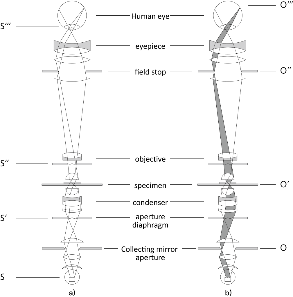
Necessary conditions for Kohler lighting:
- The light source filament is imaged on the focal plane of the condenser, illuminating the observation area of the sample with parallel light.
- The aperture diaphragm of the illumination light source is imaged on the surface of the specimen to control the size of the object’s field of view.
- Match the illumination beam to the numerical aperture of the objective lens by adjusting the field diaphragm of the illumination system.
- The aperture diaphragm and field diaphragm can be operated independently.
This illumination method is commonly used in metallographic microscopes. It has the following advantages:
- The light is similar to natural self-colored light.
- Illumination evenly.
- There are aperture diaphragms and field diaphragms that eliminate harmful light rays such as halos.
- No resolution reduction.
- Easy to operate.
Due to the above advantages of this type of lighting method, it is most suitable for microphotography, especially high-magnification microphotography.
diffuse lighting
This method sets a light diffusion plate on the reflector to achieve uniform illumination, but the brightness is weakened after passing through the light diffusion plate.
7. Types and characteristics of light sources
01 Natural light (ambient light)
External light enters the reflector to illuminate the sample. Direct sunlight must be avoided. Artificial light sources such as fluorescent lamps can also be used instead of natural light.
02 Low voltage tungsten lamp
The filament is composed of tungsten filament and is sealed with argon gas. Commonly used ones are 6V 15W and 12V 30W. It is widely used to illuminate biological samples with good transmittance, so it is mostly used in biological microscopes and teaching demonstration student microscopes. It is basically not used for metallographic microscopes that mainly observe reflective samples.
03 Halogen lamp
Halogen lamps are brighter than auto-incandescent lamps, their spectrum is close to daylight, and their color temperature changes very little over time. At the same time, it is small in size, generates less heat, and has high luminous brightness per unit area. It is a common light source for current microscopes, especially metallographic microscopes. Generally, 12V 100W is used.
04 Xenon lamp
The xenon lamp uses the inert gas xenon as the luminous element, and is commonly used as a short arc xenon lamp. Its spectrum is close to the human eye, with high brightness, excellent light color quality, and a color temperature of about 6000k. Xenon lamps are widely used in high-speed and color photography, large-field projection and television observation. Since the xenon lamp requires a starting device when ignited, the structure is complex and the volume is large, so it is more expensive. At present, metallographic microscopes are basically not used.
05 Ultra-high pressure mercury lamp
This is a point source high intensity UV lamp. It is suitable for fluorescence observation. The spectrum is a linear peak spectrum. A high-voltage starting device is required for ignition. Commonly used ones are 50W and 100W. The lifespan is about 200 hours. Now there is a new long-life ultra-high-pressure mercury lamp with a power of 120W and a nominal life of 2000. Hour.
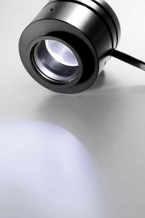
06 LED light source
Since the light emitted by LEDs has a narrow band, white light can be obtained by combining LEDs with fluorescent materials, or by combining multiple LEDs. Depending on how the LEDs are combined, LED lights are less powerful at specific wavelengths.
LEDs have many advantages over traditional lighting:
- Brightness exceeds 100w halogen lamp;
- ·High contrast, sharp image;
- ·Constant color temperature 4500K, true color reproduction;
- ·Longer service life ≥72,000 hours;
- ·Easy to adjust, no need to replace the bulb;
- ·Suitable for a variety of observation methods.