An electron microscope is an instrument that uses electron beams and electron lenses instead of light beams and optical lenses to image the fine structures of substances at very high magnifications based on the principle of electron optics.
In recent years, the research and manufacture of electron microscopes have made great progress: On the one hand, the resolution of electron microscopes has been continuously improved. The point resolution of transmission electron microscopes has reached 0.2-0.3nm, and the lattice resolution has reached about 0.1nm. , People have been able to directly observe the atomic image; on the other hand, in addition to the transmission electron microscope, a variety of electron microscopes have also been developed, such as scanning electron microscopes and analytical electron microscopes.

Although the resolving power of the electron microscope is far better than that of the optical microscope, it is difficult to observe living organisms because the electron microscope needs to work under vacuum conditions, and the irradiation of the electron beam will also cause the biological samples to be damaged by radiation. Next, the editor will talk to you about the relevant content of the electron microscope, including the principle, structure, shortcomings, application fields, and the difference between the electron microscope and the optical microscope, and its application in agriculture.
Principles of Electron Microscopy
At present, electron microscope technology has become an important means to study the microstructure of the body. Commonly used are transmission electron microscopes and scanning electron microscopes. The following describes the working principles of the two electron microscopes:
1. Transmission electron microscope
Transmission Electron Microscope commonly referred to as an electron microscope or electron microscope (EM), is the most widely used type of electron microscope.
- Working principle: Under vacuum conditions, after the electron beam is accelerated by high voltage, it forms scattered electrons and transmitted electrons when it penetrates the sample, and they are imaged on the fluorescent screen under the action of the electromagnetic lens. When the electron beam is projected onto the sample, corresponding electron emission can occur according to the density of the tissue components. For example, when the electron beam is projected onto a structure with a large mass, the electrons are scattered more, so the electrons projected on the fluorescent screen are less and appear dark. Like, electronic photos are black.
- Main advantages: high resolution, can be used to observe the ultrastructure inside tissues and cells as well as the whole picture of microorganisms and biological macromolecules.
2. Scanning Electron Microscope
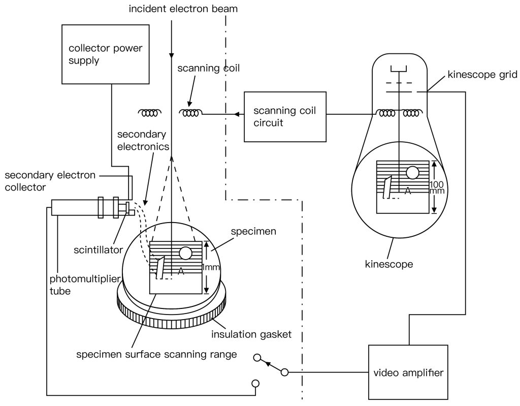
A scanning electron microscope is a scanning electron microscope, which is mainly used to observe the surface morphology of the sample, the structure of the split surface, and the structure of the inner surface of the lumen.
- Working principle: SEM uses secondary electron signal imaging to observe the surface morphology of samples. Scan the surface of the sample with a very fine electron beam, excite the surface of the sample to emit secondary electrons, collect the generated secondary electrons with a special detector, form an electrical signal and send it to the picture tube, and display the object on the fluorescent screen. The three-dimensional conformation of the (cell, tissue) surface can be photographed.
- Main advantages: the depth of field is long, and the images obtained have a strong sense of three-dimensionality, which can be used to observe various morphological features of biological samples.
Three main structures of an electron microscope
The electron microscope consists of three parts: electron optical system, vacuum system and power supply system.
The following three parts are introduced respectively:
1. Electron optical system
- The electron optical system mainly includes electron guns, electron lenses, sample holders, fluorescent screens and camera mechanisms, etc. These components are usually assembled into a cylinder from top to bottom.
- The electron gun is composed of a tungsten hot cathode, grid and cathode. It can emit and form an electron beam with a uniform speed, so the stability of the accelerating voltage is required to be no less than one ten-thousandth.
- The electron lens is the most important part of the electron microscope lens barrel. It uses a space electric field or magnetic field symmetrical to the axis of the lens barrel to bend the electron track to the axis to form a focus. Its function is similar to that of a glass convex lens to focus the beam, which is called an electronic lens. Most modern electron microscopes use electromagnetic lenses, focusing electrons through a strong magnetic field generated by a very stable DC excitation current passing through a coil with pole shoes.
2. Vacuum system
In order to ensure that it only interacts with the sample in the entire channel and does not collide with air molecules, therefore, the entire electron channel from the electron gun to the photographic backplane box must be placed in a vacuum system, generally with a vacuum degree of 10.4 to 10.7 mm Hg.
3. Power supply system
The transmission electron microscope needs two parts of power supply: one is to supply the high voltage part of the electron gun, and the other is to supply the low voltage steady current part of the electromagnetic lens. The stability of the power supply is an extremely important sign of the performance of the electron microscope. Therefore, the main requirement for the power supply system is to generate high and stable accelerating voltage and excitation current for each lens. In addition to the above-mentioned power supply parts, modern instruments also have automatic operation program control systems and computer systems for data processing.
Six Advantages of Electron Microscopes
- Magnification and higher resolution – since it uses electrons rather than light waves, it can be used to analyze structures that would otherwise not be visible. Electron microscope images have a resolution of up to 0.2 nm, which is 1000 times finer than light microscopy.
- Greater depth of field – Electron microscopes have a greater depth of field, which means more specimens can be brought into focus at the same time. This feature is especially useful when inspecting samples with uneven surfaces or varying thicknesses.
- Diversified Applications – Electron microscopes have a wide range of applications in many different fields of research, including technology, industry, biomedicine, and chemistry. Application examples include semiconductor inspection, computer chip fabrication, quality control and assurance, atomic structure analysis, and drug development.
- High-quality images – With proper training, electron microscope operators can use the system to generate highly detailed, high-quality structural images, revealing complex and delicate structures that may be difficult to reproduce with other techniques.
- Elemental Analysis – Electron microscopes can be equipped with Energy Dispersive X-ray Spectroscopy (EDS) or Electron Energy Loss Spectroscopy (EELS) systems, enabling elemental analysis of samples. This capability is invaluable in materials science, geology and other fields where identifying and mapping chemical elements is critical.
- Transmission electron microscopes are often used to observe the fine material structures that cannot be resolved by ordinary microscopes; scanning electron microscopes are mainly used to observe the morphology of solid surfaces, and can also be combined with X-ray diffractometers or electron energy spectrometers. Constitute electron microprobes for material composition analysis; emission electron microscopes are used for the study of self-emitting electron surfaces.
Eight Limitations of Electron Microscopy
- 1. In the electron microscope, the sample must be observed in a vacuum, so it is impossible to observe the live sample. With the advancement of technology, the environmental scanning electron microscope will gradually realize the direct observation of the live sample;
- 2. When processing the sample, it may produce a structure that the sample does not have, which exacerbates the difficulty of analyzing the image afterward;
- 3. Due to the strong electron scattering ability, secondary diffraction is easy to occur;
- 4. Because it is a two-dimensional plane projection image of a three-dimensional object, sometimes the image is not unique;
- 5. Since the transmission electron microscope can only observe very thin samples, it is possible that the structure of the surface of the material is different from the structure inside the material;
- 6. For ultra-thin samples (less than 100 nanometers), the sample preparation process is complicated and difficult, and the sample preparation is damaged;
- 7. The electron beam may destroy the sample through collision and heating;
- 8. In addition, the purchase and maintenance prices of electron microscopes are relatively high.