1. What is a phase contrast microscope
A phase contrast microscope is a common microscope, which can be seen in many laboratories. It uses the principle that the phase difference generated when light passes through a transparent sample is converted into the amplitude or contrast difference in the image. It is mainly used to observe bacteria, cells transparent samples of cellular structures. Because it can examine living cells in a natural state without prior killing, fixing, and staining of cells, phase contrast microscopy can observe and record dynamic biological processes with high definition and high contrast, and can be used for cell cycle, etc. direction of research.
The emergence of phase contrast microscopy benefits from the invention of phase contrast microscopy, which is a major advancement in microscopy technology, and its inventor Zernike won the 1953 Nobel Prize in Physics for this.
2, The principle of phase contrast microscopy
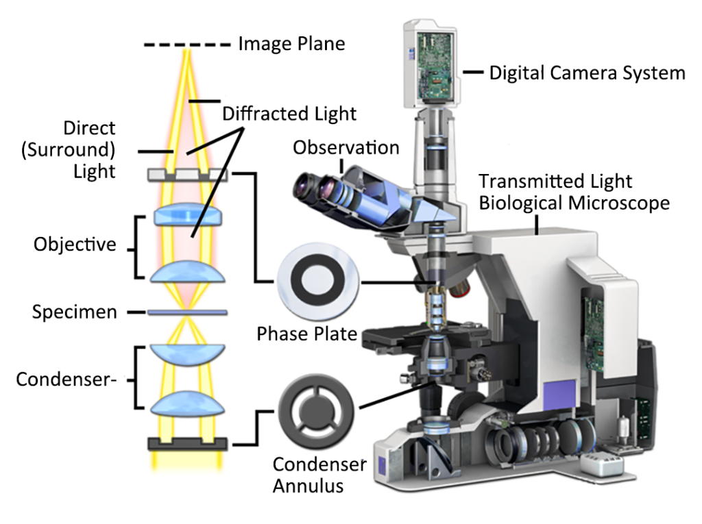
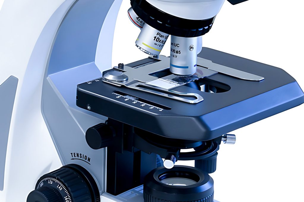
The core of the phase contrast microscope is a phase contrast ring plate located at the aperture stop of the condenser and a phase plate located behind the objective lens. First, the light is emitted from a point in the filament for illumination, and is precisely focused on the opening of the phase difference ring plate at the condenser by the field lens. Since this position is in the front focal plane of the condenser, the light will become parallel light after passing through the condenser. Assuming that the two beams of light do not diffract at the specimen, they will enter the objective lens in parallel, and all the parallel light rays will be focused on the phase plate behind the objective lens. But in fact, part of the light will be diffracted after passing through the specimen. In order to complete the phase adjustment requirement, it is necessary to adjust the phase plate behind the objective lens. Another function of the phase plate is to attenuate the light that is not affected by the specimen. Use a centering telescope to observe the phase plate behind the objective lens, and you can see the dark ring formed by the light-reducing material.
The phase plate behind the objective lens is composed of a conjugate surface and a compensation surface. The conjugate surface is ring-shaped and is the part through which direct light passes. It is usually coated with light-absorbing substances. light part.
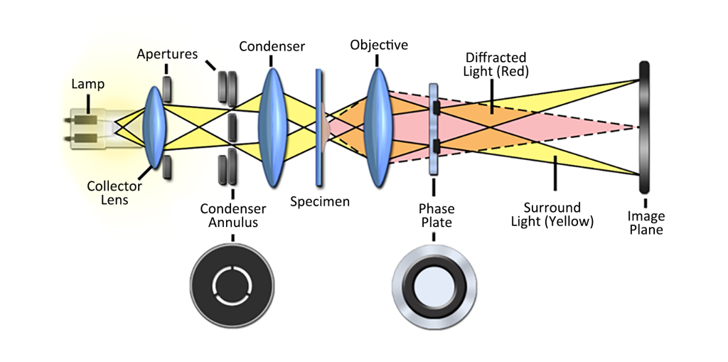
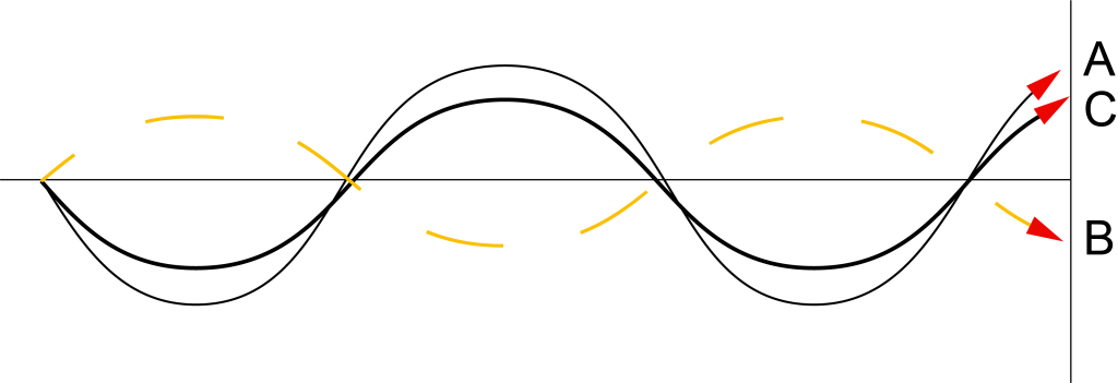
The light enters through the phase difference ring plate and exits through the condenser to form a parallel wave front. When these plane waves hit the sample, some of the light is diffracted as it passes through the sample, changing the phase relationship between them and the direct light, and the phase of the newly generated spherical wave will be delayed by λ/4 after exiting the sample. Note that there are now two types of waves, direct light and diffracted light, with a phase difference of λ/4 between them. In positive phase contrast, the conjugate surface is only coated with an absorption film, and the compensation surface is coated with a phase film, so that the direct light A is only absorbed, the amplitude is reduced, and the phase is not delayed; while the phase of the diffracted light B is delayed by λ/ 4. The phase difference between the direct light and the direct light is λ/2 (λ/4+λ/4), and the amplitude of the synthetic wave C on the intermediate phase plane is equal to the difference between the amplitudes of the two waves (causing destructive interference). At this time, the sample It is darker than the background (because the background only has direct light, which is absorbed and the amplitude is reduced), which is the effect of positive phase contrast.
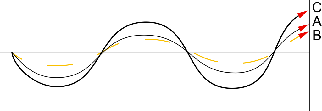
On the contrary, in negative phase contrast, since the conjugate surface of the phase plate is not only coated with an absorbing film, but also coated with a phase film, in addition to absorbing the direct light to reduce its amplitude, it also delays the phase of the direct light by λ/4, In this way, the direct light A and the diffracted light B maintain the same phase, and the synthetic wave C is equal to the sum of the amplitudes of the direct wave and the diffracted wave (causing constructive interference), because the background is only illuminated by the direct light, and the sample is brighter than the background.
In this way, the phase difference of direct light or diffracted light is changed through the phase contrast microscope, and the phase difference that cannot be distinguished by the human eye becomes the difference in the amplitude of light energy intensity (light and dark difference) that the eye can measure reaching the retina, and it will not be observed when observing cells. Harm to cell specimens.
3. Advantages of Phase Contrast Microscopy
Phase contrast microscopy can observe samples such as unstained living cells. Compared with other contrast-enhancing techniques, such as DIC, phase contrast microscopy does not use polarized light illumination, so it can observe birefringent media, such as living cells through plastic culture bottles or dishes. Observation, the internal structure of the sample (such as the nucleus) can be clearly observed, which is especially useful in cytological research. If it is used in conjunction with epifluorescence technology, the phase contrast objective lens can transmit about 70% of the excitation light, which is better than DIC, so phase contrast is often used as an auxiliary imaging function in fluorescence microscopes. In addition, the purchase price of the phase difference kit is relatively low, and the adjustment is relatively simple.
However, phase contrast microscopy has some inherent shortcomings. Phase-contrast imaging has low axial resolution due to the necessity to use an annular diaphragm and the limited numerical aperture of the condenser when phase-contrast illumination is enabled. In addition, obvious halos will appear at the interface where the refractive index difference between the specimen and the background is large, resulting in the obscuration of details such as cell membranes and outer edges of organelles.
4. Application of Phase Contrast Microscopy
Phase contrast microscopy is primarily used for high-contrast imaging of transparent specimens such as living cells (usually in culture), thin tissue sections, photolithographic patterns, fibers, latex dispersions, glass fragments, and subcellular particles (including nuclei and other organelles ).
In addition, the phase contrast microscope is widely used in clinical practice, especially for the inspection of cells and microorganisms in fresh liquid specimens, especially for live cells. Generally, a fresh liquid sample is taken and a cover glass is added (for example, when using an oil lens, oil must be added to the cover glass) for immediate inspection. Such as: blood platelet count, blood parasite inspection and some live cell observation in hematology test; rapid identification of unstained or unstained bacteria, spirochetes, fungal spores, etc. in microbiology test, especially It is the identification of microorganisms with special morphology such as flagella and capsule, and it is also used in urinalysis to identify various cells and casts in fresh urine, especially the classification of hyaline casts, white blood cells, renal tubular epithelial cells, and red blood cell shapes, etc.