1. What is an optical microscope?
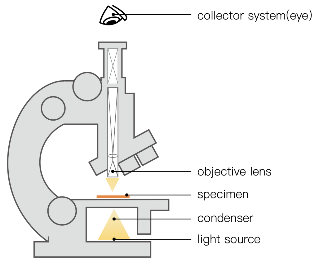
An optical microscope ( Light microscope) is a microscope that uses optical lenses to produce image magnification effects.
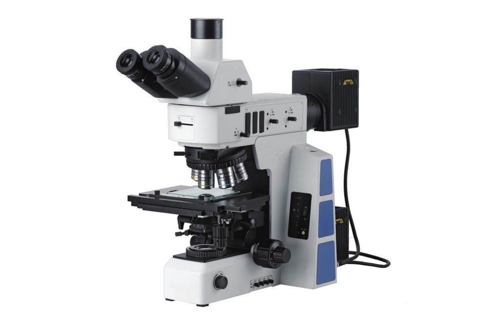
Light incident by an object is amplified by at least two optical systems (objectives and eyepieces). First, the objective lens produces a magnified real image, and the human eye observes this magnified real image through the eyepiece, which acts like a magnifying glass. A general optical microscope has multiple replaceable objective lenses, so that the observer can change the magnification as needed, that is, increase the magnification. The magnification is obtained by multiplying the eyepiece magnification by the objective lens magnification. These objective lenses are generally placed on a rotatable objective lens disk. By rotating the objective lens disk, different objective lenses can easily enter the optical path. The English name of the objective lens disk is Nosepiece, also translated as nose wheel.
In the eighteenth century, the magnification of optical microscopes had been increased to 1,000 times, allowing people to see clearly the shape, size, and some internal structures of microorganisms with their eyes. It was not until physicists discovered the law between magnification and resolution that people knew that the resolution of optical microscopes has a limit. This limit of resolution limits the infinite increase in magnification. 1600 times becomes the magnification of optical microscopes. The highest limit of magnification greatly limits the application of morphology in many fields. The resolution of an optical microscope is limited by the wavelength of light, which generally does not exceed 0.3 microns. Resolution can also be improved if the microscope uses ultraviolet light as a light source or if the object is placed in oil.
Optical microscopes can be divided into reflection and transmission types according to different samples. The objects in reflection microscopes are generally opaque, light shines on the object from above, and the light reflected by the object enters the microscope. This kind of microscope is often used to observe solids, etc., and is mostly used in the fields of engineering and materials. Among upright microscopes, this type of microscope is also called a metallographic microscope. A transmission microscope object is transparent or so thin that light can pass through it and enter the microscope. This type of microscope is often used to observe biological tissue.
An optical microscope can be used to observe different samples depending on the design of its condenser and objective. Brightfield is used to observe thin stained biological tissue samples. Under the darkfield function, the background is black, which can highlight the subtle features of the sample. When observing unstained samples, such as living cells, the phase difference can be used (Phase) function. There is also a differential interference contrast (DIC) function, which is often used with optical microscopes.
Depending on the light source, there are also categories such as fluorescence microscopes and confocal microscopes.
2. What is an electron microscope?
An electron microscope is a microscope that uses electrons to display the interior or surface of an object.
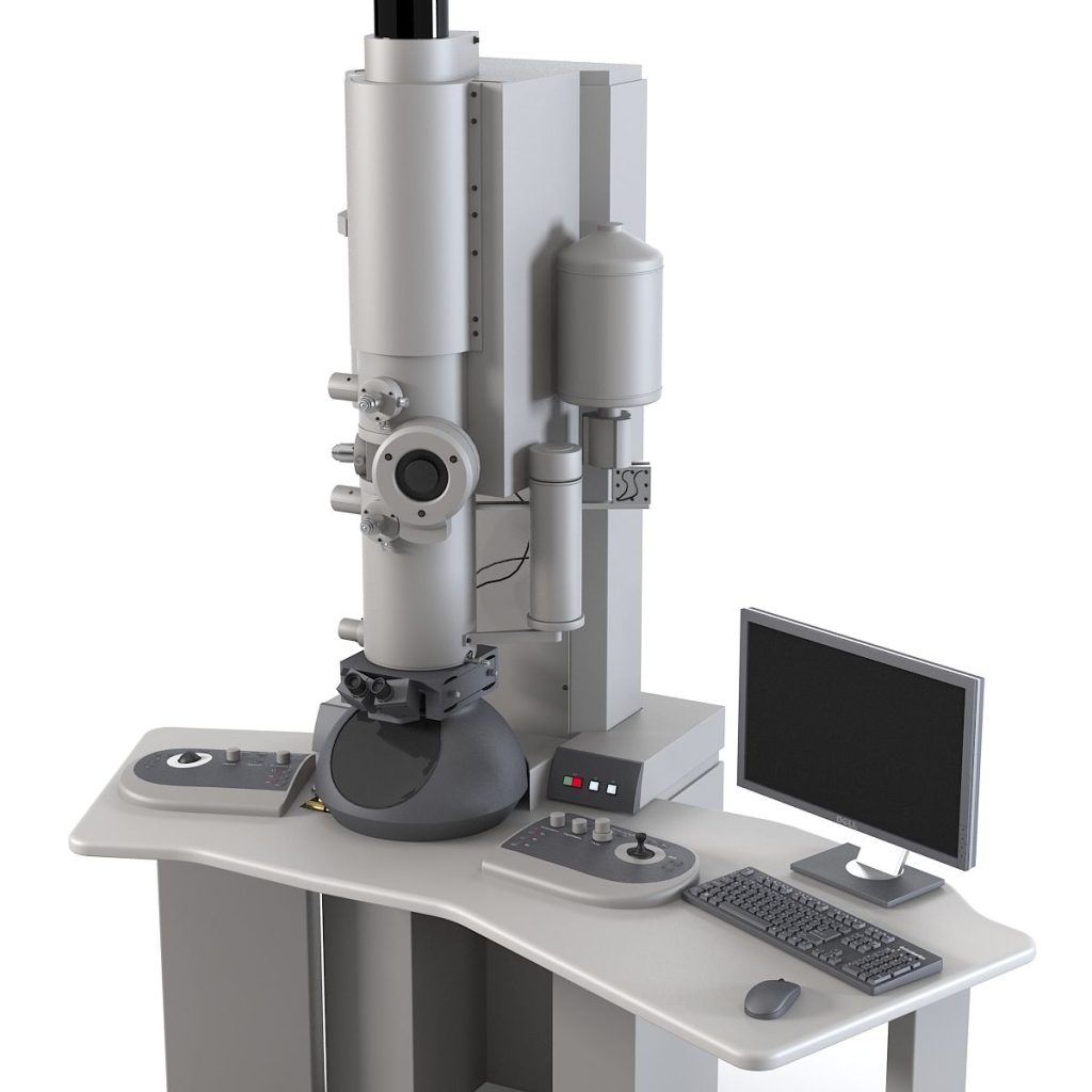
The wavelength of high-speed electrons is shorter than the wavelength of visible light (wave-particle duality), and the resolution of a microscope is limited by the wavelength used. Therefore, the resolution of an electron microscope (about 0.2 nanometers) is much higher than that of an optical microscope. rate (approximately 200 nm).
Technology
The main components of an electron microscope are:
The electron source is a cathode that releases free electrons, and a ring-shaped anode accelerates the electrons. The voltage difference between the cathode and anode must be very high, typically between a few thousand volts and 3 million volts.
Electron lenses, are used to focus electrons. Generally, magnetic lenses are used, and sometimes electrostatic lenses are also used. The function of the electron lens is the same as that of the optical lens in an optical microscope. The focus of the optical lens is fixed, while the focus of the electron lens can be adjusted, so the electron microscope does not have a movable lens system like the optical microscope.
Vacuum device. The vacuum device is used to maintain a vacuum within the microscope so that electrons are not absorbed or deflected along their path.
Sample holder. Samples can be placed stably on the sample holder. In addition, there are often devices that can be used to change the sample (such as moving, rotating, heating, cooling, stretching, etc.).
The detector is used to collect electronic signals or secondary signals.
3. Seven differences between optical microscopes and electron microscopes
- 1. Different imaging principles
The basic principle of optical microscopy is to use the different characteristics of the different structures of the sample to absorb light to present the object image of the sample in the form of brightness difference. In an electron microscope, a finely focused electron beam is used to scan the sample surface point by point, interacting with the sample to produce various physical signals. These signals are received, amplified and converted into modulated signals by the detector, and finally displayed on the fluorescent screen to reflect the sample surface. Images with various features.
- 2. Different lighting sources
The illumination source of an optical microscope is visible light (sunlight or light), while the illumination source used in an electron microscope is the electron stream emitted by an electron gun. Since the wavelength of the electron stream is much shorter than the wavelength of light, the magnification and resolution of the electron microscope are significantly higher than those of the light microscope. .
transmission electron microscope
- 3. Different lenses
The objective lens that plays a magnifying role in an electron microscope is an electromagnetic lens (a ring-shaped electromagnetic coil that can generate a magnetic field in the center), while the objective lens in an optical microscope is an optical lens made of ground glass. There are three groups of electromagnetic lenses in electron microscopes, which have the same functions as a condenser, objective lens and eyepiece in optical microscopes.
- 4. Depth of field
Generally, the depth of field of an optical microscope is between 2-3um, so it has extremely high requirements for the surface smoothness of the sample, so the sample preparation process is relatively complicated. The depth of field of the scanning electron microscope can be as high as several millimeters, so there is no requirement for the smoothness of the sample surface. Sample preparation is relatively simple, and some sample geometries do not require sample preparation. Although stereo microscopes also have a relatively large depth of field, their resolution is very low.

- 5. Different preparation methods of specimens used
The preparation procedures for tissue and cell specimens used for electron microscopy observation are complex, technically difficult, and expensive. Special reagents and operations are required in aspects such as material extraction, fixation, dehydration, and embedding. The embedded tissue blocks also need to be placed Human ultramicrotome cuts ultra-thin specimen slices with a thickness of 50~100nm. Specimens for light microscopy observation are generally placed on glass slides, such as ordinary tissue section specimens, cell smear specimens, tissue-pressed specimens, and cell drop specimens.
- 6. Resolution
Due to the interference and diffraction of light, the resolution of optical microscopes can only be limited to 2-5um. Because the electron microscope uses electron beams as the light source, its resolution can reach between 1-3nm. Therefore, the tissue observation under the optical microscope belongs to micron-level analysis, while the tissue observation under the electron microscope belongs to nano-level analysis.
- 7. Application fields
Optical microscopes are mainly used for the observation and measurement of micron-level structures on smooth surfaces. Because visible light is used as the light source, not only the surface tissue of the sample can be observed, but also the tissue within a certain range below the surface layer can also be observed, and the optical microscope is very sensitive to color. The recognition is very sensitive and accurate. Electron microscopes are mainly used to observe the surface morphology of nanoscale samples. Because SEM relies on the intensity of physical signals to distinguish tissue information, the images of SEM are all black and white, and SEM is powerless to identify color images.