Introduce
Atomic Force Microscopy (AFM) was developed to overcome a fundamental shortcoming of Scanning Tunneling Microscopy (STM) – it can only image conductive or semiconductor surfaces. The strength of atomic force microscopy is that it can image virtually any type of surface, including polymers, ceramics, composites, glasses, and biological samples.
Atomic force microscopy (AFM) has high-resolution imaging features at the nanometer or even sub-nanometer scale. It is designed to meet the growing demand for imaging, analysis, and characterization of nanoscale features.
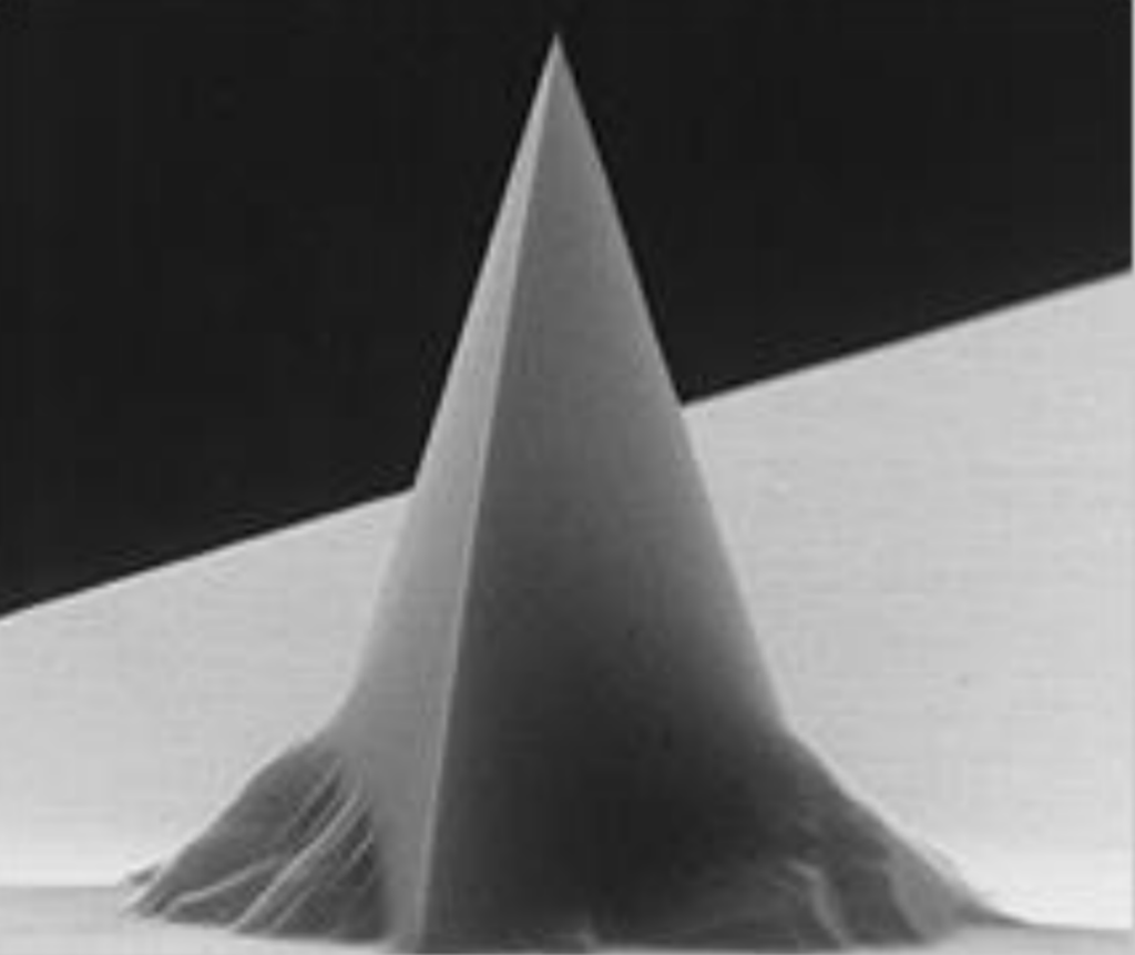
An atomic force microscope consists of a cantilever with a very sharp tip (<10nm) at the end to scan the surface of the sample. The tip material is usually doped silicon, but can also be made of other materials with higher stiffness and hardness such as SiN, SiC, and diamond.
Depending on the scan mode, the AFM measures contact forces (contact mode) or van der Waals forces (non-contact mode) by deflecting the cantilever by bringing the tip close to the sample surface, since the surface has features and waviness of different heights, the AFM cantilever The degree of deflection also changes. Thus, 3D surface topography can be analyzed based on AFM cantilever deflection.
Imaging is not the only function that AFM can provide, the system can also be used for the measurement of many other properties, such as nanomechanical properties, electrical properties, magnetic properties, and chemical/biological properties.
AFM Operating Modes
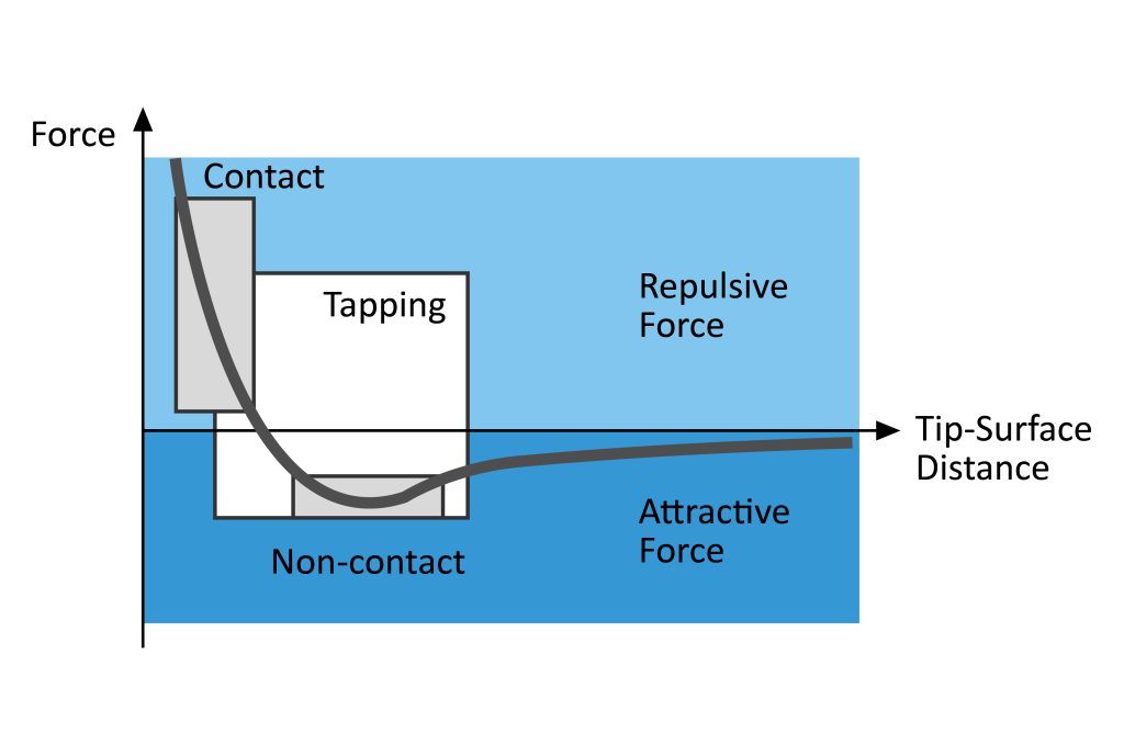
There are two basic modes of imaging surface topography using AFM: static or contact mode and dynamic mode. According to the interaction force between the AFM tip and the surface, the dynamic mode is further subdivided into tapping or intermittent contact and non-contact modes.
1. In contact mode, the AFM probe tip scans the entire sample surface while maintaining constant contact with the sample surface (Figure 2). The feedback system is designed to maintain a constant AFM cantilever deflection and thus a constant interaction force. The force between the AFM tip and the surface is repulsive (Figure 3). Soft AFM cantilevers with force constants ≤1N/m are generally used to minimize AFM tip wear and surface damage and to improve sensitivity.
One of the major disadvantages of contact mode operation is that the AFM tip on a soft AFM cantilever is susceptible to lateral forces and will adhere to surface contamination layers present on most surfaces in ambient air. These can cause image distortion. Additionally, lateral forces can damage AFM probe tips and soft samples.
2. In tapping mode, the AFM probe cantilever is oscillated by a piezoelectric actuator at or near its fundamental resonant frequency, typically tens to hundreds of kilohertz. The AFM probe is lowered towards the sample surface so that the AFM probe tip lightly touches the surface at the lower end of the AFM cantilever oscillation (Figure 4), whose oscillation amplitude is damped. A feedback loop maintains a constant AFM cantilever oscillation amplitude and therefore a constant interaction force. The forces that dominate the interaction between the AFM tip and the surface are repulsive forces (Figure 3).
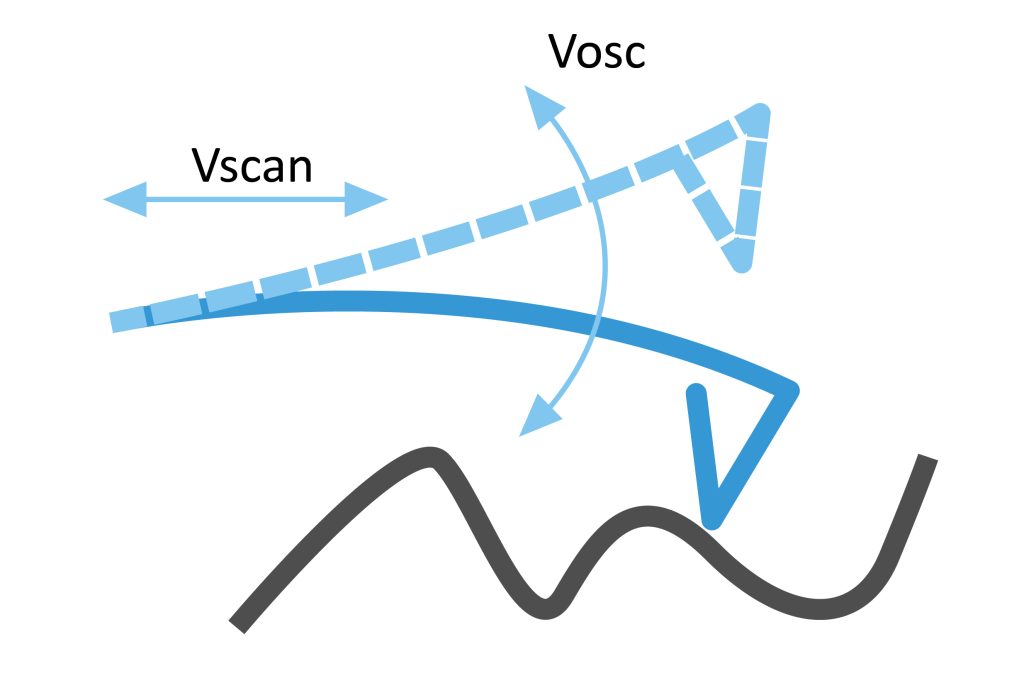
Rigid AFM cantilevers with force constants in the 10-100N/m range and high resonance frequencies >190kHz for standard tapping mode operation. These AFM cantilevers will not stick to surfaces when measuring in ambient air. For measurements on soft samples, such as living cells, softer AFM cantilevers with force constants <10N/m and sometimes as low as sub-0.1N/m are used.
3. In the non-contact mode, the AFM cantilever oscillates at or near its resonant frequency with a smaller amplitude than in the tapping mode (1nm or less), and the AFM probe tip is kept at a distance of a few nanometers to tens of nanometers from the surface (Fig. 5) in the region of attractive interaction forces (Fig. 3). In some cases, a feedback loop maintains a constant AFM cantilever oscillation frequency and therefore a constant interaction force (FM-AFM). This approach could provide more precise force control and ultra-high-resolution images in liquids.
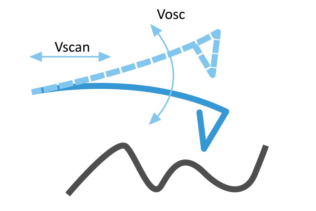
The advantage of the non-contact mode is that it provides the lowest possible interaction between the tip and the sample surface. Small interaction forces help maintain AFM tip sharpness and achieve high resolution. The downside is that it can be challenging to keep the AFM tip in an attractive range. For small tip-surface distances, high-performance feedback control is required.
AFM cantilevers with high force constants and high resonance frequencies are best suited for non-contact mode.
Atomic Force Microscopy Imaging Procedure
Atomic force microscopy imaging includes three steps: surface sensing, detection method and imaging:
1. Surface Sensing
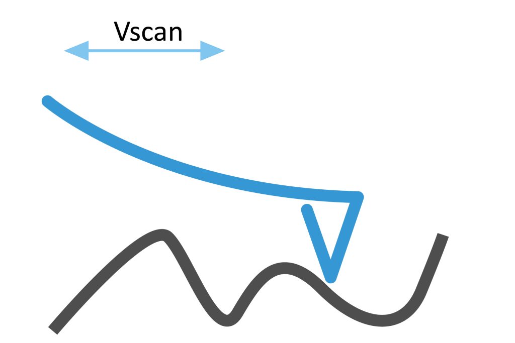
An atomic force microscope uses a cantilever with a very sharp tip to scan the surface of a sample. As the tip approaches the surface, the close-range attractive force between the surface and the tip causes the cantilever to deflect toward the surface.
However, as the cantilever is brought closer to the surface, such that the tip makes contact with it, more and more repulsive forces take over and cause the cantilever to deflect away from the surface.
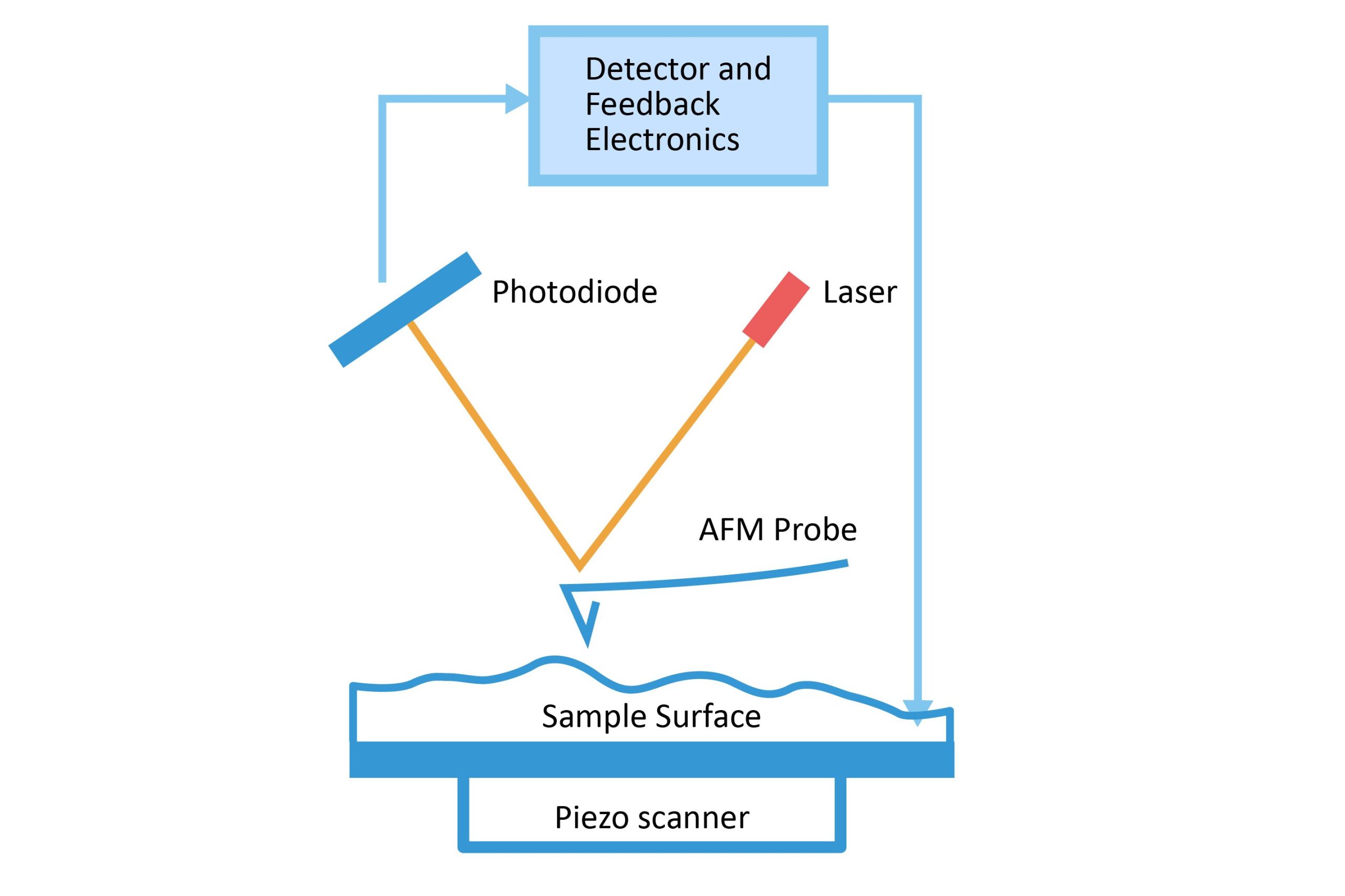
2. Detection method
A laser beam is used to detect the deflection of the cantilever towards or away from the surface. By reflecting the incident beam from the flat top of the cantilever, any cantilever deflection causes a slight change in the direction of the reflected beam.
Position-sensitive photodiodes (PSPDs) can be used to track these changes. Thus, if the AFM tip passes over a raised surface feature, the resulting deflection of the cantilever (and subsequent change in direction of the reflected beam) is recorded by the PSPD.
3. Imaging
An atomic force microscope images the topography of a sample surface by scanning a cantilever over a region of interest. Raised and lowered features of the sample surface affect the deflection of the cantilever, which is monitored by the PSPD.
By using a feedback loop to control the height of the tip above the surface—thus maintaining a constant laser position—the atomic force microscope can generate precise topographic maps of surface features.
Resolution of Atomic force microscopy
The higher the resolution of the AFM image, the more information can be extracted about the smallest features on the sample surface.
Vertical (Z-axis) imaging resolution is probe independent. On the other hand, the lateral resolution obtained is usually limited by the tip geometry of the AFM probe rather than the capabilities of the microscope.
Mid-level AFMs are typically capable of sub-nanometer lateral resolution and sub-Angstrom vertical resolution. High-quality standard AFM probes have sub-10 nm AFM tip radii and allow low nanometer resolution.
Atomic or near-atomic resolution at the atomic plane can be achieved with high-resolution AFM probes with tip radii in the range of 1-5 nm.
Of course, high resolution can only be achieved if the original extreme sharpness of the AFM tip is preserved during the measurement by scanning under optimal conditions that limit AFM tip wear and AFM tip contamination.
Advantages of Atomic force microscopy
AFM has many advantages over optical microscopes/profilometers and scanning electron microscopes (SEM).
Atomic force microscopy (AFM) has several advantages over scanning electron microscopy (SEM). Unlike an electron microscope, which provides a two-dimensional projection or image of a sample, an atomic force microscope provides a three-dimensional surface profile. Furthermore, AFM-observed samples do not require any special treatments (such as metal/carbon coatings) that would irreversibly alter or damage the sample.
While electron microscopes require an expensive vacuum environment to function properly, most AFM modes work flawlessly in ambient air or even liquid environments. This makes it possible to study biological macromolecules and even living organisms.
In principle, atomic force microscopy can provide higher resolution than scanning electron microscopy (SEM). It has been shown to provide true atomic resolution in ultra-high vacuum (UHV) and more recently liquid environments. High-resolution AFM’s resolution is comparable to that of scanning tunneling microscopes and transmission electron microscopes.