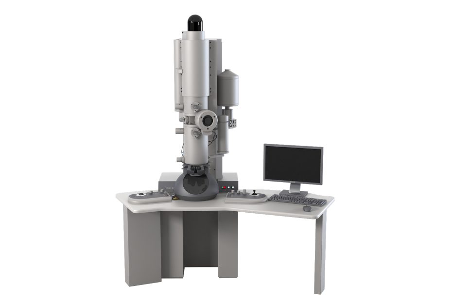
Electron microscopes are important experimental equipment in scientific research laboratories, so what types of electron microscopes are there?
In fact, there are three common types: transmission electron microscope, scanning electron microscope, and scanning transmission electron microscope.
There are three types of electron microscopes:
Transmission Electron Microscope (TEM), Scanning Electron Microscope (SEM), Scanning Transmission Electron Microscope (STEM)
1. Transmission electron microscope (TEM)
The sample must be made into a film that is transparent to electrons and has a thickness of 100-2000Å. The imaging method is similar to the optical biological microscope, except that the glass lens is replaced by an electronic lens. The magnified electronic image is displayed on the fluorescent screen.
Figure 1 is a schematic diagram of the optical path of a transmission electron microscope. The resolution of TEM can reach about 3Å. It can be higher in special cases.
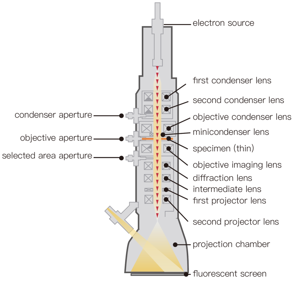
(1) Ultra-high voltage electron microscopy (HVEM) is a kind of TEM, but the commonly used TEM acceleration voltage is 100kV, which can only penetrate samples with a thickness of several thousand angstroms. HVEM is similar to general TEM except for the acceleration tube, but the size is enlarged. The 1000kV electron microscope is two stories high.
After the size is enlarged, the space around the sample increases, which is convenient for placing various accessories for processing samples, such as stretching, heating, cooling, chemical reaction, and other accessories, and can combine with the inclined sample stage; it can also do dynamic observation, Changes during sample handling were recorded with a TV.
High-energy electrons can cause radiation damage in samples, which brings great convenience to the study of the microscopic mechanism of material radiation damage.
(2) The high-resolution electron microscope (HREM) increases the accelerating voltage to make the electron wavelength shorter, which can improve the resolving power.
Due to the high technical difficulty, until the early 1970s, the ultra-high voltage electron microscope was mainly aimed at improving the penetration rate.
From the late 1970s to the early 1980s, technological improvements brought 200kV and 300kV high-resolution commercial electron microscopes and individual 500kV, 600kV, and 1000kV HREMs. The resolving power can reach about 2Å. 1.5Å will soon be possible.
Since biological molecules are easily damaged by radiation, HREM is currently mainly used to observe the arrangement of atoms in inorganic materials.
2. Scanning Electron Microscope (SEM)
It is mainly used to directly observe the morphology of the solid surface, and its principle is shown in the schematic diagram of the scanning electron microscope in Figure 2.
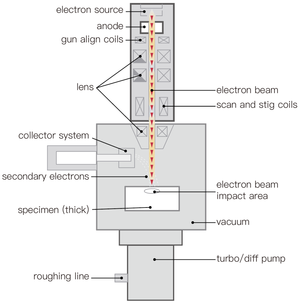
First, the electron lens is used to reduce an electron beam spot to tens of angstroms, and the deflection system is used to make the electron beam raster scan on the sample surface. The electron beam excites secondary electrons where it goes, and becomes a signal after being collected by the detector, modulating the brightness of a synchronous scanning picture tube to display the image.
The asperities on the sample surface direct some localities toward the secondary electron detector and others toward the detector. The secondary electrons emitted from the part facing the detector are more collected and appear brighter, and vice versa, appear darker, thus producing a yin and yang side, full of three-dimensional images.
The magnification of the image is the scanning amplitude of the picture tube compared to the scanning amplitude of the electron beam on the sample surface. The resolving power of SEM is slightly larger than the electron beam spot diameter. At present, the resolving power of SEM can reach 60Å.
3. Scanning transmission electron microscope (STEM)
The imaging method is similar to the scanning electron microscope, but instead of receiving secondary electrons, it receives transmitted electrons (including some small-angle scattered electrons). The sample must also be a thin film, and the resolving power of STEM is equivalent to the diameter of the electron beam spot.
Special high-brightness field emission electron gun for STEM (requires ultra-high vacuum of 10-10 Torr). The resolving power can reach 3Å. Single heavy atoms on light element support membranes have been observed using this STEM.
It is particularly important for practical work that its tiny electron beam spot can be used for crystal structure analysis (using electron diffraction) and composition analysis (using electron beam excited X-rays or electron energy loss) in extremely small areas (tens of angstroms). Spectrum).
At present, TEM can be equipped with STEM accessories, but because there is no high-brightness field emission gun, the beam spot can only be reduced to tens of angstroms. Capable of structural and compositional analysis in the range of about 100 Å.
An electron microscope that can perform the above-mentioned analysis on any tiny part of it while observing the microscopic image is called an “analytical electron microscope”.
TEM maintenance:
1. Always turn on the machine and use it more often, so that you can grasp the working conditions of the instrument at any time, pay attention to the changes in pictures, light, sound, vacuum, air pressure, and power supply, adjust them in time, and make good records. After a long time, you will definitely accumulate a lot of experience.
2. Pay attention to air humidity, prevent rats from spilling, stable voltage, clean and dry gas, prevent small samples from falling in, especially fine particles, and powder, and prevent collisions
3. Monitor whether the freon in the electron gun is lowered, monitor whether the oil in the mechanical pump is lowered below the horizontal line, ventilate the air compressor frequently, replace the water in the circulating water device regularly, and keep the indoor humidity at 40~70%. The temperature is between 15 and 25 degrees.
For the repair of electron microscopes, it is necessary to have a high technical level, practical ability, and the flexible application of superb comprehensive knowledge. At the same time, it also depends on the accumulation of experience. It is best to be someone who has studied electronics and has practical ability. To manage the electron microscope.
4. The vacuum oil used in the rotary pump should be changed once a year if possible. The speed of oil contamination is mainly related to the cleanliness and humidity of the laboratory. It is best to turn on the electron microscope when it is not in use. Vacuum every day, and the circuit is energized every other day or two. It is best to turn it on two days a week when the school is on holiday.