The emergence of the scanning electron microscope provides new methods and means for human beings to explore the mysteries of the microcosm. Compared with optical microscopes, scanning electron microscopes have the advantages of high resolution, large depth of field, less damage to samples, and light pollution. There are very broad applications in other fields.
Difference Between Electron Microscope and Light Microscope Compare the Difference Between Similar Terms
1. Different lighting sources:
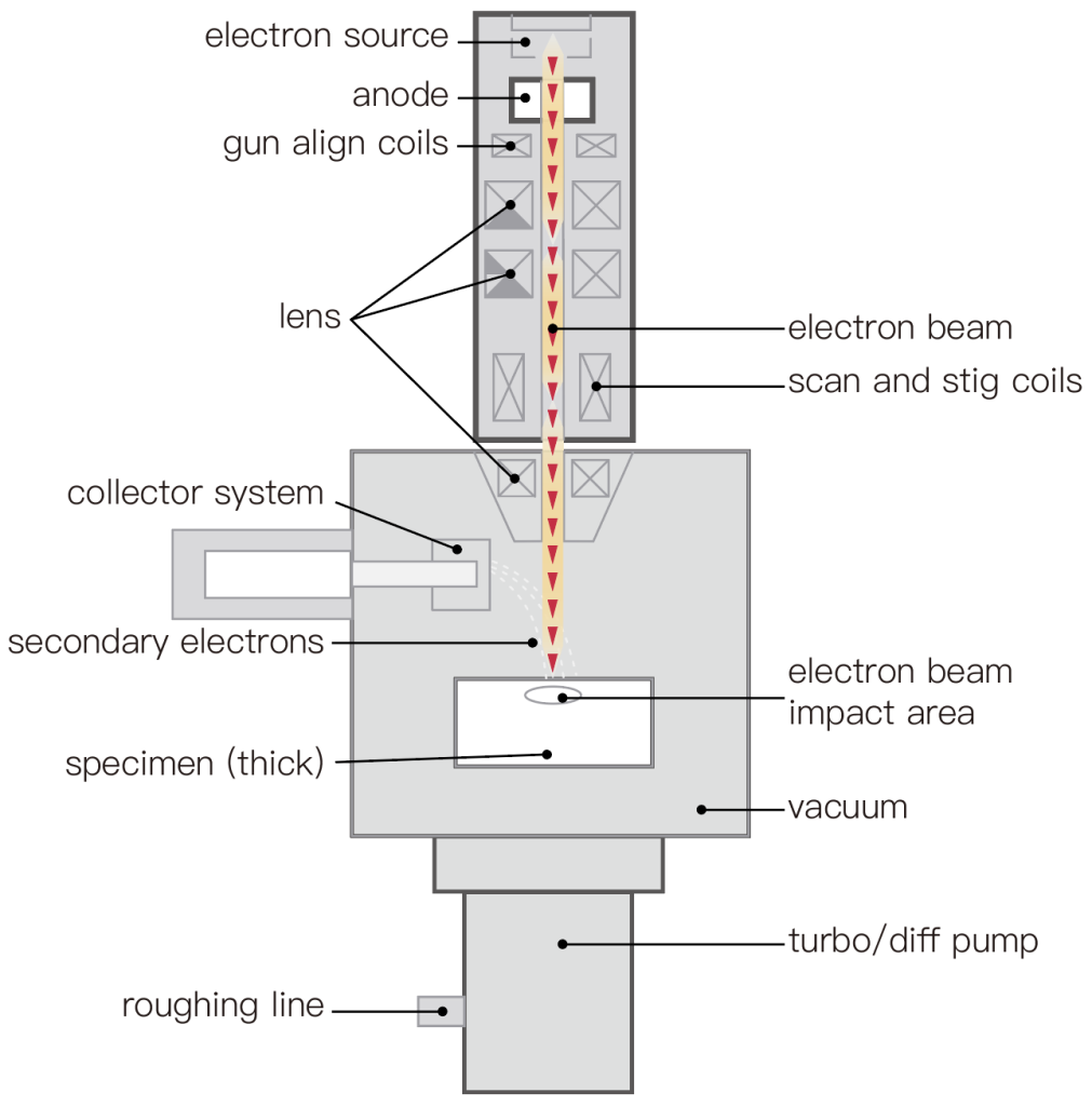
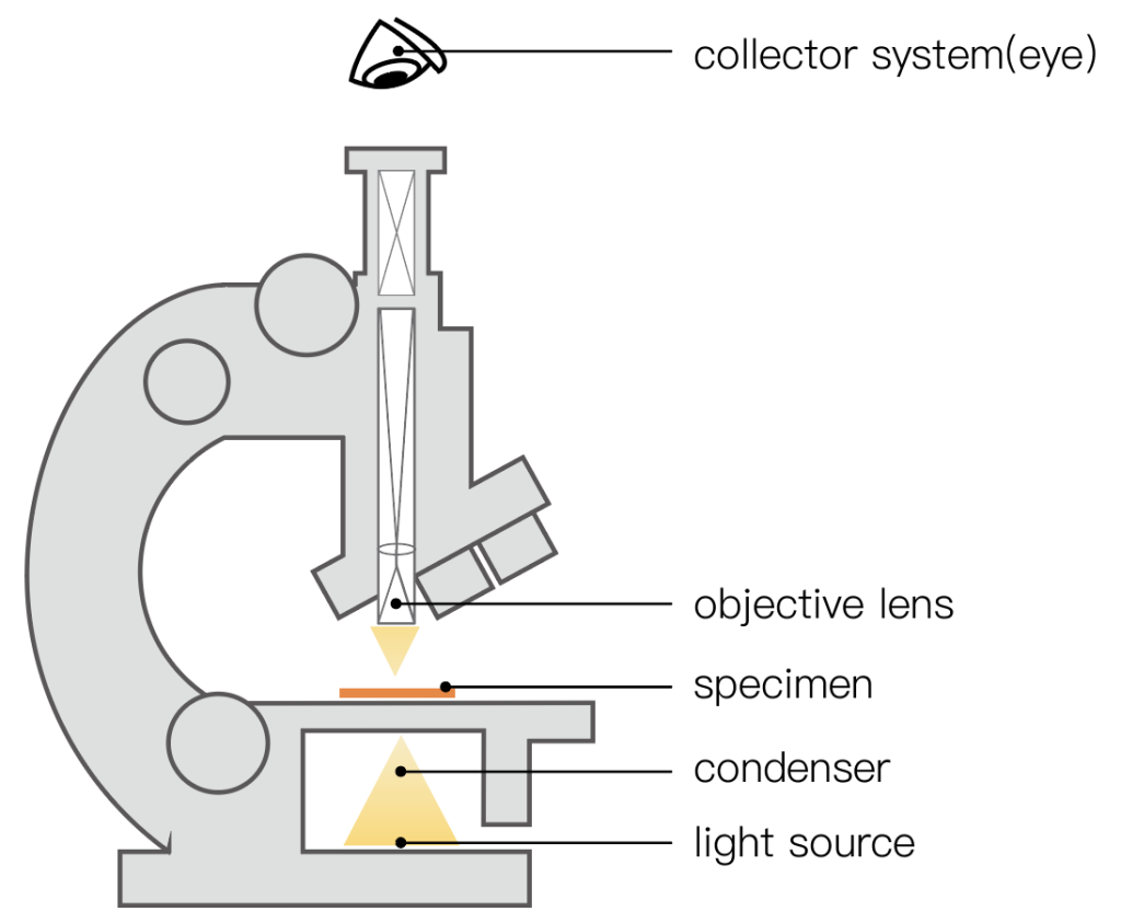
The illumination source used by the electron microscope is the electron flow emitted by the electron gun, while the illumination source of the light microscope is visible light (sunlight or light). Since the wavelength of the electron flow is much shorter than that of the light wave, the magnification and resolution of the electron microscope are significantly higher than that of the light microscope.
2. The lens is different:
The magnifying objective lens in the electron microscope is an electromagnetic lens (an annular electromagnetic coil that can generate a magnetic field in the central part), while the objective lens of the light mirror is an optical lens made of glass. There are three groups of electromagnetic lenses in the electron microscope, which are equivalent to the functions of the condenser lens, objective lens and eyepiece in the light microscope.
3. Different imaging principles:
In the electron microscope, the electron beam acting on the sample to be inspected is magnified by the electromagnetic lens and hits the fluorescent screen for imaging or acts on the photosensitive film for imaging. The mechanism of the difference in electron density is that when the electron beam acts on the sample to be tested, the incident electrons collide with the atoms of the substance to produce scattering. Since different parts of the sample have different scattering degrees for electrons, the electron image of the sample is presented in shades. The object image of the sample in the light microscope is presented as a difference in brightness, which is caused by the difference in the amount of light absorbed by the different structures of the sample under inspection.
4. Resolution:
Due to the interference and diffraction of light, the resolution of optical microscope can only be limited to 02-05um. Because the electron microscope uses electron beams as the light source, its resolution can reach between 1-3nm. Therefore, the tissue observation of the optical microscope belongs to micron-scale analysis, and the tissue observation of electron microscope belongs to nano-scale analysis.
5. Depth of field:
Generally, the depth of field of an optical microscope is between 2-3um, so it has extremely high requirements on the smoothness of the surface of the sample, so the sample preparation process is relatively complicated. The spirit of the scanning electron microscope can be as high as several millimeters, so there is no requirement for the smoothness of the sample surface geometry, the sample preparation is relatively simple, and some sample geometry does not require sample preparation. Although the stereo microscope also has a relatively large depth of field, its resolution is very low. Magnification: The effective magnification of the optical microscope is 1000X. Electron microscope effective magnification.
6. The specimen preparation methods used are different:
The preparation procedure of tissue cell specimens used for electron microscope observation is complicated, and the technical difficulty and cost are high. Special reagents and operations are required in the links of material collection, fixation, dehydration and embedding. Finally, the embedded tissue blocks need to be put into Cut into ultra-thin specimen slices with a thickness of 50-100nm by a human ultra-microtome. The specimens observed by light microscopy are generally placed on glass slides, such as ordinary tissue section specimens, cell smear specimens, tissue compression specimens and cell drop specimens. It can reach 1000,000X.
This article focuses on the application of scanning electron microscopy in the field of biology, and analyzes it from the aspects of palynology, diatom inspection, and biomedicine.
Application of Scanning Electron Microscope in Biological Field
(1) Palynology
There are many kinds of plants in nature, and there are also many flowering plants. The pollen of different types of plants will have unique and stable morphological characteristics, but the pollen morphology of plants of the same genus often have high similarity, so palynology research has great significance in plant taxonomy. important reference value.
There are certain differences in pollen morphology among different species. Using scanning electron microscope to observe the size of pollen shape, germination groove and surface decoration of outer wall has important taxonomic significance for the division of species.
The surface morphology, shape, and details of the pollen can be clearly seen through the scanning electron microscope. For example, the lotus pollen is a three-groove pollen under the scanning electron microscope, which is ellipsoidal in shape and has fine thorn-like protrusions on the surface; Liliaceae leaf pollen The grains are boat-shaped, with a single germination groove at the far pole; the pollen shape of lemon germplasm is nearly spherical, prolate and oblate; These are things that optical microscopes cannot do.
Using scanning electron microscopy to clarify the morphological differences of plant pollen can lay a certain theoretical foundation for the study of plant phylogenetic evolution, classification and phylogenetic relationship.
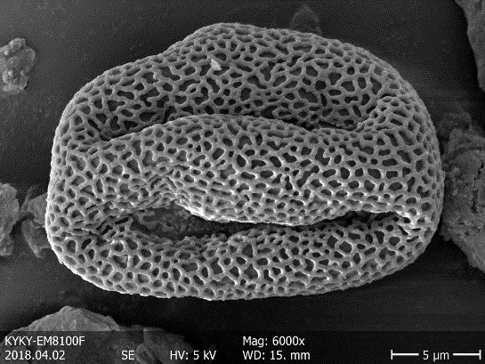
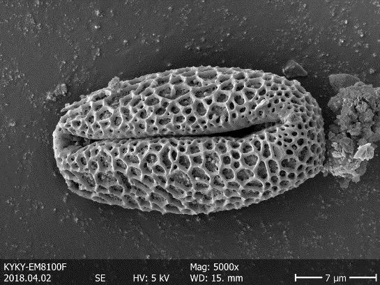
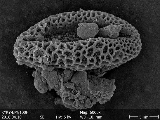
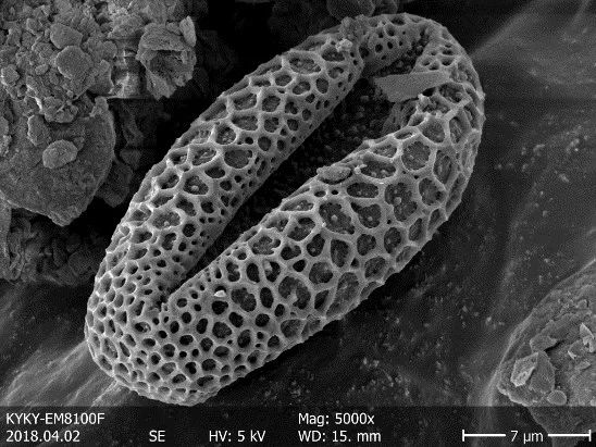
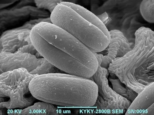
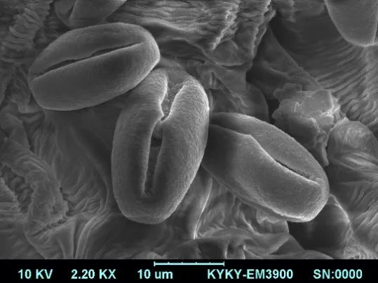
Figure 1: Pollen
(2) Diatom inspection
The scanning electron microscope has excellent performances such as high resolution, large depth of field, and non-destructive to the sample, and can be used in conjunction with an X-ray energy spectrometer, so that it can quickly conduct a comprehensive analysis of the surface morphology and micro-area composition of tiny substances, so as to provide Forensic science provides objective basis and clues for criminal investigation.
The use of scanning electron microscopes to inspect and identify human tissue, hair, plant fragments, diatoms, pollen, insects, microorganisms and other organisms will not destroy the original form of physical evidence, and can be used to judge the time of death and provide clues for case analysis.
Diatom detection method is of great significance in forensic drowning diagnosis. Observing the shape and type of diatoms through scanning electron microscope can find out the location of the water body. If abnormal types of diatoms are found on the clothes, it can indicate that the corpse has been moved.
In addition, scanning electron microscopy can also be used to inspect a wide variety of trace evidence such as shooting residues, glass, car paint, pollen, rubber, plastic, fiber, hair, metal, soil, etc. that appear at the scene of the case, which is a must for criminal investigation and forensic science. indispensable means of analysis.
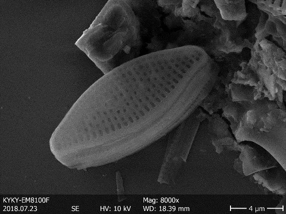
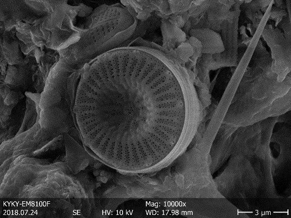
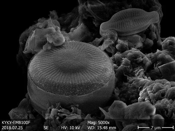
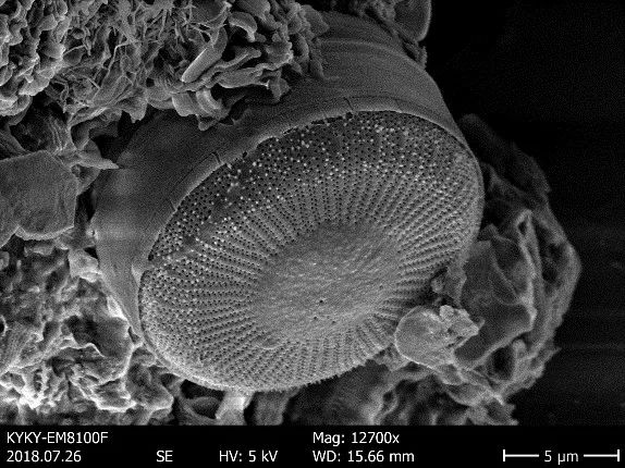
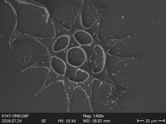

Figure 2: Diatoms
(3) Biomedicine
The microscopic structure of materials determines the macroscopic properties of materials, and it is very necessary for medical workers to master microscopic information. The application of scanning electron microscopy technology can analyze the structure and chemical composition of surgical implants from the micro-nano scale, and analyze the cell structure, cell substructure, cell ultrastructure and virus structure, which is indispensable for surgical implantation and pathological analysis. lack of research tools.
The scanning electron microscope combined with the energy spectrometer can not only obtain the structure and topography of the sample, but also perform three-dimensional reconstruction at the micro-nano scale, and can give corresponding chemical information and elemental analysis on the sample surface. For example, using scanning electron microscopy to observe the three-dimensional structural changes of lung tissue, especially the morphological changes of alveolar structure, is a new way to study the mechanism of highly pathogenic invasion of lung tissue. The atypical cancer cells under the scanning electron microscope showed dendritic shape, rough surface, and protrusions of various shapes, and the cell volume was large. According to the ultrastructural changes of cells, combined with biological characteristics such as molecular expression and immune activity of cells, it can provide strong and beneficial help for the research of cancer prevention, targeted drugs, and therapeutic methods.
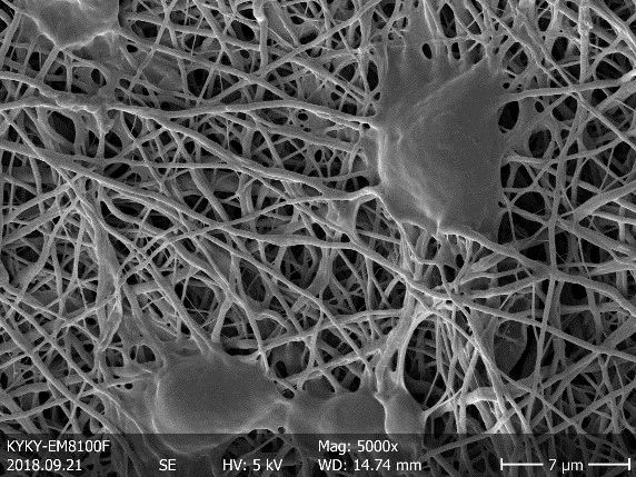
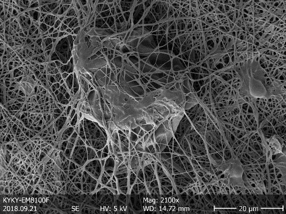
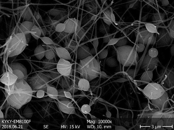
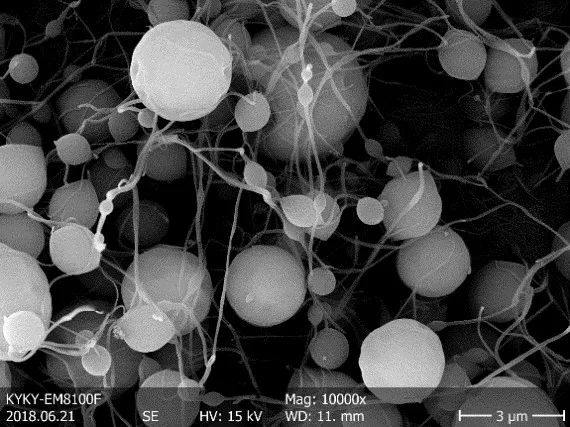
Figure 3: Cells
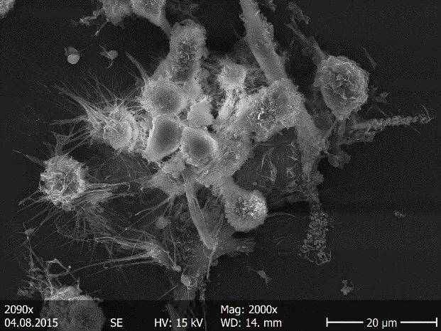
Figure 4: Viruses
Summarize
Scanning electron microscopes are essential instruments for cutting-edge scientific research. All the above observations are taken with a scanning electron microscope to help users conduct biological research more intuitively, conveniently and intelligently.
In recent years, the company has successively launched field emission gun series scanning electron microscopes, whose performance indicators have reached the international advanced level, and have been widely used in materials science, life science, aerospace, environmental protection, semiconductor manufacturing and other fields.
At the same time, actively respond to national policies and market demand, develop a variety of high-end instruments, and provide customized solutions for scientific research and industrial customers!