There are three main signals in the imaging process of SEM: secondary electrons, backscattered electrons, and characteristic X-rays.
Secondary electrons (SE) and backscattered electrons (BSE) are the two most basic and commonly used signals in scanning electron microscopy (SEM). For many SEM users, secondary electrons can be used to characterize the morphology. It is common knowledge that scattered electrons can be characterized by atomic number.
Secondary Electronics
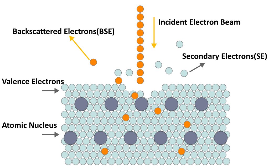
Secondary electrons are commonly known as “morphological images”. Under the impact of incident electron beams on the surface atoms of the sample, the extranuclear electrons of the sample atoms are bombarded out, and the extranuclear electrons that leave the sample surface are called secondary electrons, as shown in Figure 1.
Secondary electrons are generally emitted within the depth range of 5-10nm in the surface layer, which is very sensitive to the surface topography of the sample, so it can display the surface topography of the sample very effectively.
Since it is a low-energy electron, the energy is lower than 50 eV, and the energy of most secondary electrons is lower than 10 eV, the secondary electrons generated in the depth range of 50~500Å have the opportunity to escape from the surface of the sample and be detected.
The characteristics of secondary electrons are as follows:
(1) Since the energy is less than 50eV, it is easier to be attracted by the electric field at the front end of the detector, so the shadow effect is weaker.
(2) Only the secondary electrons excited by the very shallow part of the sample surface (about 10nm) can escape the sample surface, so the resolution of the secondary electron image is high;
(3) The output of secondary electrons mainly depends on the local slope of the sample surface, so the secondary electron image is mainly a topographic image. It can be seen as consisting of details such as convex points, steps, and pits formed by many surfaces with different inclinations. Different parts of these details emit different numbers of secondary electrons, resulting in contrast.
The secondary electron image has a high resolution, no obvious shadow effect, a large depth of field, and a strong stereoscopic effect. It is the main imaging method of the scanning electron microscope and is especially suitable for the observation of the surface morphology of rough samples.
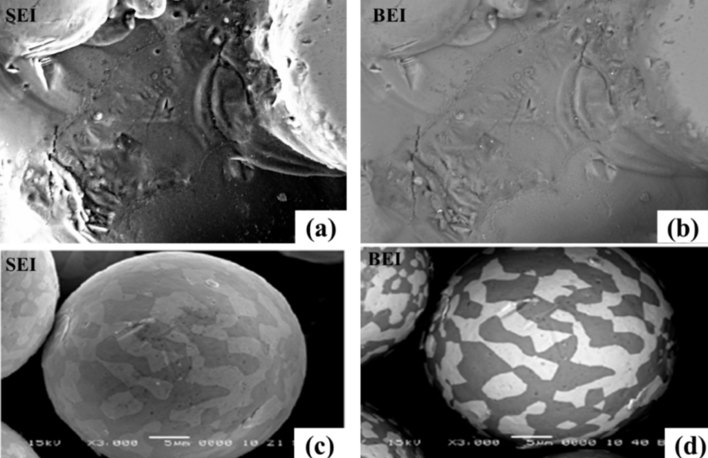
As shown in (a) of Figure 2, it can be clearly found from the SEI image that the surface of the sample is uneven, and the darker parts are pits or cracks.
Backscattered Electrons
Backscattered electrons, commonly known as “atomic number images”, are high-energy electrons produced by the elastic or inelastic scattering of incident electron beams and atomic nuclei.
The yield of backscattered electrons (BSE), that is, the ratio of the number of outgoing backscattered electrons (BSE) to the number of incident electrons, depends on the average atomic number of the sample: the higher the average atomic number, or the heavier the element, the brighter the contrast.
As shown in Figure 1. Its kinetic energy is equal to or slightly less than the energy of incident electrons, and the excitation area is larger than that of secondary electrons.
The characteristics of backscattered electrons are as follows:
(1) The energy is high, and the penetration ability is much higher than that of secondary electrons, from 50eV to close to the energy of incident electrons.
(2) The penetrating ability is much stronger than that of secondary electrons and can escape from a deeper area in the sample (micron level). In such a depth range, the incident electrons have a fairly wide lateral expansion, so in the sample The generated range is large and the image resolution is low;
(3) The amount of backscattered electrons increases significantly with the increase of the atomic number. The parts with higher atomic numbers backscatter more electrons and form brighter areas on the screen; while the parts with lower average atomic numbers produce more electrons. Fewer backscattered electrons create darker areas on the phosphor screen, which creates atomic number contrast (compositional contrast).
(4) Strictly speaking, backscattered electrons also carry shape information, but it is not as obvious as secondary electrons.
As shown in (b) of Figure 2, it can be found from the BEI image that there is little difference in the composition of this region, and the reflection of the topographic features of the BEI image is inferior to that of the SEI image. For the comparison of (c) and (d) in Figure 2, it can be clearly seen that the BEI image has the advantage of observing the composition distribution, and the difference of different tissue components in the powder can be clearly seen.