Two common illumination methods are used in microscopy: “transmitted illumination” and “incident illumination.” “Transmitted illumination” is suitable for transparent or semi-transparent objects under examination, with the vast majority of biological microscopes falling into this category. On the other hand, “incident illumination” is suitable for non-transparent objects, with the light source coming from above, also known as “reflected illumination,” primarily used in metallographic microscopes or fluorescence microscopy.
This article discusses the choice between the two illumination methods in microscopy.
Transmitted Illumination
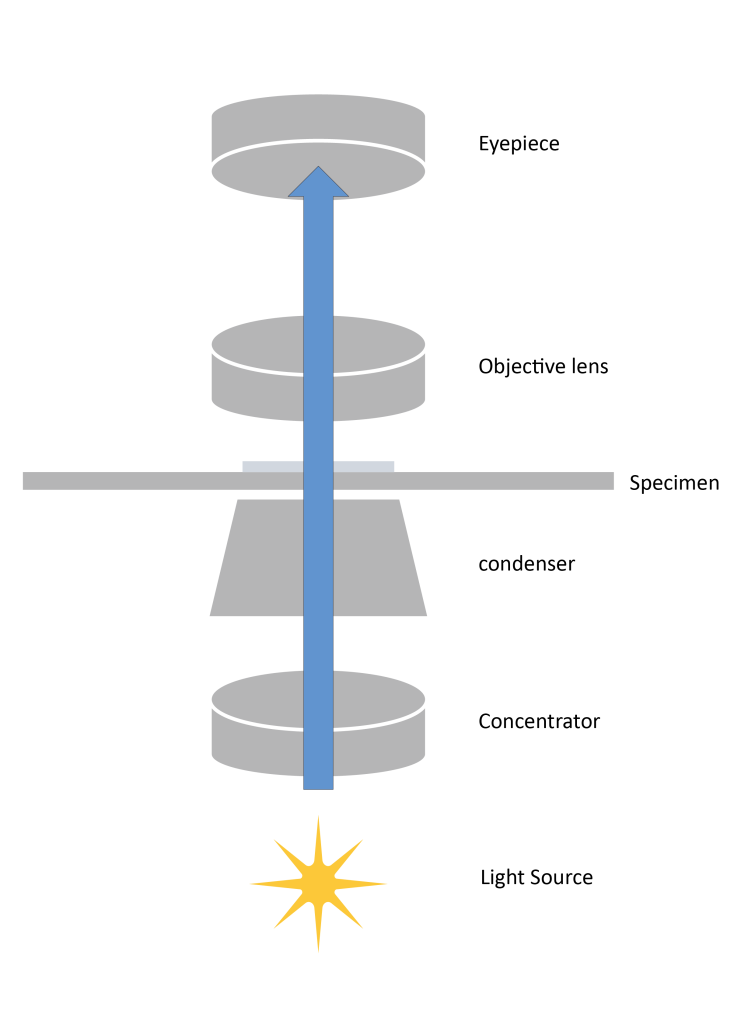
From the optical path diagram, it can be seen that the light source passes through the condenser, aperture diaphragm, focusing lens, sample, objective lens, and eyepiece, and finally enters our field of view.
Transmitted illumination can be divided into “critical illumination” and “Köhler illumination.”
A. Critical Illumination
The characteristic of this illumination is that the light source, after passing through the focusing lens, forms a filament image on the object being examined. The light beam is narrow and intense, which is its advantage. However, the filament image of the light source and the object being examined are in the same focal plane, which can interfere with imaging.
B. Köhler Illumination
Köhler illumination overcomes the drawbacks of critical illumination. This illumination method provides excellent observation results and is essential for microscopic photography.
The advantages of Köhler illumination are primarily as follows:
- The filament does not fall on the plane of the examined object, resulting in uniform illumination.
- The thermal focal point of illumination is not on the object being examined, preventing damage to it.
- The focusing lens images the field diaphragm onto the plane of the object being examined, and adjusting the size can control the illumination area.
Incident Illumination
Incident illumination is when the light beam comes from above the object, passing through the objective lens and then onto the object being examined, where the objective lens also acts as a condenser. This illumination method is suitable for observing non-transparent objects, such as metals and minerals.
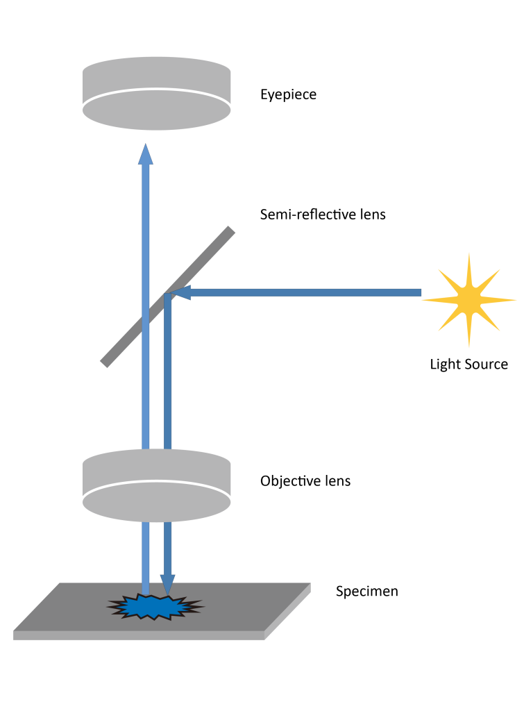
In the microscope’s incident light structure, a vertical illuminator is added between the observation head and the body, with a semi-reflective lens at the intersection of the vertical illuminator and the objective lens’s optical path. From the optical path diagram above, we can see that after passing through the semi-reflective lens, 50% of the light reflects onto the sample, while the other 50% passes through the semi-reflective lens. The sample then reflects the light source and passes through the semi-reflective lens again, with finally 25% of the light passing through and entering the eyepiece.
Furthermore, while the incident light in fluorescence microscopy and metallographic microscopy follow a similar optical path, their principles are entirely different. In incident fluorescence microscopy, there is a dichroic mirror in the optical path, which functions based on the wavelength of light to determine whether it undergoes total reflection or total transmission: short wavelengths undergo total reflection, while long wavelengths undergo total transmission.
The main advantage of incident illumination is that the illuminated area and the observed area are nearly identical, avoiding stray light entering the field of view, resulting in uniform illumination and superior imaging quality. Additionally, incident illumination imposes higher requirements on the objective lens; besides magnification, the objective lens must also function as a condenser, leading to higher-quality lenses for better imaging results.