The iSTORM ultra-high-resolution microscopy imaging system uses STORM technology derived from the Nobel Prize in Chemistry principles to achieve 20nm spatial resolution. It provides imaging tools for analysis of subcellular structures such as vesicles, positioning and counting of biological macromolecules, and kinetic studies; it also provides fluorescent dye selection, sample preparation, imaging services, and overall experimental solutions to support scientific researchers in constantly challenging higher resolutions. Achieve breakthroughs in the fields of life sciences and medicine.
Super-resolution imaging
Optical microscopes have long been an important tool in biomedical research due to their non-contact and non-destructive advantages.
The resolution of ordinary optical microscopy imaging technology is sufficient for human cells with an average diameter of 10-20um.
Electron microscopes can achieve nanometer-level resolution and can observe the positioning of organelles such as vesicles and mitochondria inside cells. However, due to the lack of specific probe labels, they are not suitable for locating individual protein molecules, nor are they suitable for observing living cells and cell membranes. dynamic change process.
Therefore, biologists are eager to have an experimental microscopy method that not only has optical resolution capabilities at the submicron or even nanometer scale but also can continuously monitor the evolution of biological macromolecules and tiny structures of organelles without affecting the biological activity of biological systems. .
Super-resolution Optical Microscopy came into being. Super-resolution optical imaging breaks the resolution limit of optical microscopes (in other words, exceeds the resolution limit of optical microscopes, so it is called super-resolution optical imaging) and increases the resolution from hundreds of nanometers to tens of nanometers. Provides unprecedented tools for life science research.
There are three main types of super-resolution microscopy
There are three main types of super-resolution microscopy, each working through a different mechanism. These include stimulated emission depletion (STED) microscopy, structured illumination microscopy (SIM), and stochastic optical reconstruction microscopy (STORM)/photoactivated localization microscopy (PALM).
Super-resolution microscopy imaging technology has important applications in discovering new intracellular structures, biomolecular interactions, quantitative molecular counting, and dynamic time tracking.
Introduction to ultra-high resolution microscopy imaging system
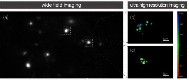
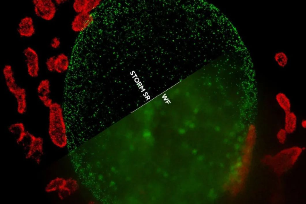
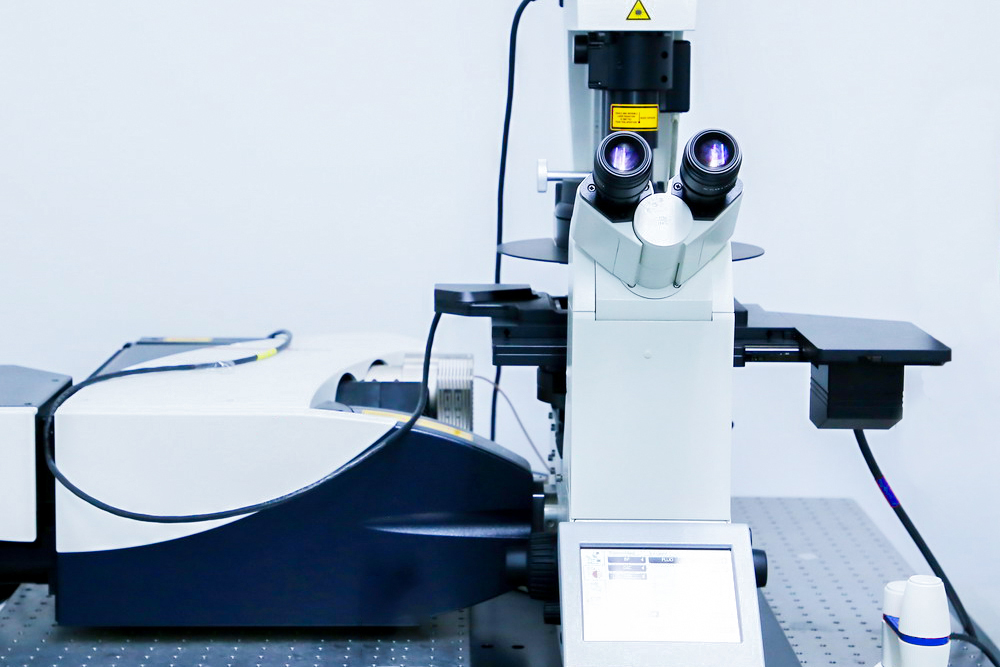
The iSTORM super-resolution microscopy imaging system is a high-end imaging equipment based on single-molecule positioning microscopy imaging technology. The complete machine includes the main body of the microscope, laser and controller system, total internal reflection illumination system, multi-channel imaging system, active locking system, living cell life support system (optional), and computer imaging workstation. iSTORM utilizes the scintillation characteristics of dyes in specific buffer environments to precisely position the position of single-molecule dyes, thereby increasing the spatial resolution of imaging from 200nm (XY direction) and 500nm (z direction) of ordinary fluorescence microscopes to 20nm. (XY direction) and 50nm (Z direction).
iSTORM can provide a variety of imaging channel configurations of dual channels, three channels, and four channels to meet various imaging needs; it can be used with a living cell life support system for ultra-high imaging of living cells, and can analyze living cells with high-precision imaging capabilities Various life and physiological processes; and the system adopts a multi-channel simultaneous imaging mode, which makes the imaging speed faster and can avoid the loss of sample structure information caused by erroneous excitation to the greatest extent; the system is equipped with an active locking system, which ensures that during the imaging process The sample position is locked at 1 nanometer to avoid the impact of sample drift on the imaging results; it has the intellectual property rights of the entire system, so the system can be expanded in any feasible way according to needs. During the development process of the system, a large number of sample shots have been accumulated With our experience, we can provide optimized sample production solutions and imaging result analysis services.
The ultra-high-resolution microscopy imaging product iSTORM, as an ultra-high-resolution microscopy system with independent intellectual property rights, is based on the 2014 Nobel Prize-winning technology by applying a series of physical principles, chemical mechanisms and algorithms. “Breakthrough” the optical diffraction limit, increasing the resolution of optical microscopes tenfold, allowing humans to observe the biological microscopic world from an unprecedented perspective below 200nm.
Features of iSTORM ultra-high resolution microscopy imaging system
- 1. Ultra-high spatial resolution – advanced technology, 20nm ultra-high imaging
(Using STORM random optical reconstruction technology and adding cylindrical mirror design, the XY-axis resolution reaches 20nm and the Z-axis resolution reaches 50nm, with 3D imaging function)
- 2. Optimized time resolution – multi-channel simultaneous imaging optical path design
It adopts a proprietary multi-channel simultaneous imaging optical path design to provide a stable optical path. The self-developed imaging spectroscopic optical path can ensure that the optical paths between channels are relatively independent, so that the fluorescence emitted by the sample can be received by the detector with maximum efficiency, minimizing crosstalk between channels. In conjunction with the optimal dye solution and optimal imaging buffer formula, multi-channel simultaneous imaging can achieve 20nm ultra-high resolution imaging within a time range of several minutes to more than ten minutes.
- 3. High system stability – real-time locking design of nanoscale physical samples
Adopting nano-level real-time dynamic locking technology, real-time physical compensation is used to correct sample drift. The locking accuracy is 1nm. No preheating is required. It is ready for use. It is easy to operate and is free from environmental influences on the sample position such as airflow, temperature changes, noise, mechanical vibration, etc. It can stabilize imaging even in high-rise buildings, noisy, vibrating, and normal temperature environments. Therefore, it has the characteristics of high efficiency, simplicity, good adaptability to the environment, and is friendly and easy to use.
- 4. Live cell imaging–supporting cutting-edge scientific research
As the basic unit and functional unit of life, cells are of great significance in exploring the essence of life through the study of the internal structure and functions of living cells. Traditional optical microscopy technology is limited by the diffraction limit and cannot observe biological structural details below 200nm. Super-resolution fluorescence microscopy technology improves traditional imaging resolution by 10-20 times by providing optical images with spatial resolution below the diffraction limit, opening up new opportunities for studying nanometer-sized biological structures. Our company has launched the iSTORM ultra-high-resolution microscope, which can be used with appropriate live-cell probes to perform ultra-high-resolution imaging of living cells, providing a powerful tool for scientific researchers to deeply study the internal molecular structure of living cells.
- 5. High scalability-customizable multi-modal function combination
The system can be equipped with functional modules such as SPT, confocal, FRET, and live cell monitoring. At the same time, the system is independently developed by our company and has a complete set of technical reserves including hardware, software, algorithms, biological sample preparation, etc., so it can meet personalized customization needs and can provide customized services such as hardware, software, and algorithms.
iSTORM super-resolution imaging in research
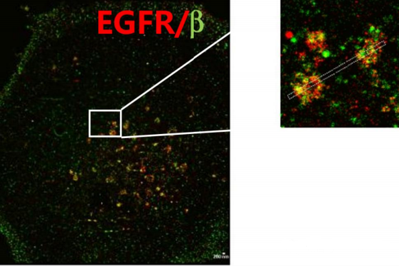
- 1. Study the functions of proteins and related applications of cell organelles;
- 2. Live cell photography;
- 3. Research on the pathogenicity mechanism of viruses;
- 4. Research on the mechanism of GPCR action in tumors and other diseases;
- 5. Related to blood diseases;
- 6. Drug research and development and treatment effect evaluation, etc.;
- 7. Nucleic acid, in situ hybridization;
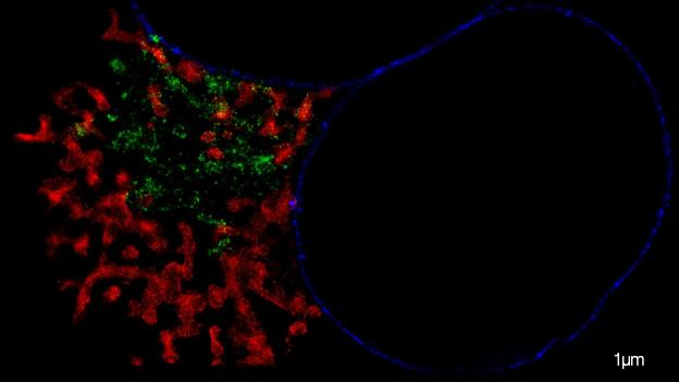
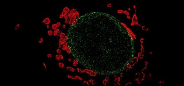
- 8. Intracellular mechanics, membrane dynamics, organelle distribution, internalization and transport processes;
- 9. Photographing plant cells, such as studying the dynamics of plant cell membrane proteins;
- 10. Research on the mechanism of action of proteins related to neurological diseases;
- 11. Study the role of exosomes in the occurrence and development of diseases;
- 12. Photographing pathological sections and studying the nanoscale tissue structure and pathogenesis of pathological aggregates;
- 13. Exploring the internalization mechanisms and transport pathways of viruses, drugs and particles;
- 14. Research on the mechanism of LLPS under the action of compounds or drugs.