The principle of fluorescence microscopy is a microscopic observation mode that combines optical microscopy with the emission of fluorescent dyes from compounds through an excitation light source. Most fluorescence microscopes used in biology today are epifluorescence microscopes.
What is fluorescence microscopy?
Fluorescence microscopes are mainly used to study samples such as organic and inorganic substances. Fluorescence and phosphorescence are generally used to examine the structural organization and spatial distribution of samples, which is more suitable for studying samples that are more complex and cannot be examined under traditional transmitted light microscopes. There are two main differences between fluorescence microscopy and traditional microscopy. One is that the type of light source is different, and the other is that the filter components used are different.
Light source (xenon or mercury arc lamp): Mercury arc lamps emit light 10-100 times brighter than most incandescent lamps and provide light in a variety of wavelengths from ultraviolet to infrared.
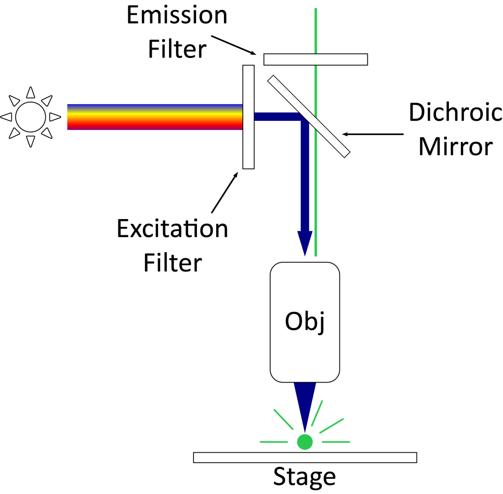
Excitation filter: The purpose of an excitation filter is to filter out all wavelengths of the light source, except the excitation range of the fluorophore being examined. The brightness and brightness of the image are determined by the minimum transmission percentage of the filter. Ideal transmittance > 85%.
A dichroic mirror (beam splitter): A dichroic mirror or beam splitter is placed at a 45° angle between the excitation filter and the emission filter. The function of the dichroic filter is to reflect the excitation signal to the fluorophore and transmit the emission signal to the detector.
Emission filter: The emission filter is located within the imaging path of the fluorescence microscope. Its job is to filter out the entire excitation range and transmit the emission range of the fluorophore being examined.
Objective Lens: The purpose of the objective lens is to transmit light onto the sample to form an image. Light passes through the dichroic mirror before reaching the objective lens.
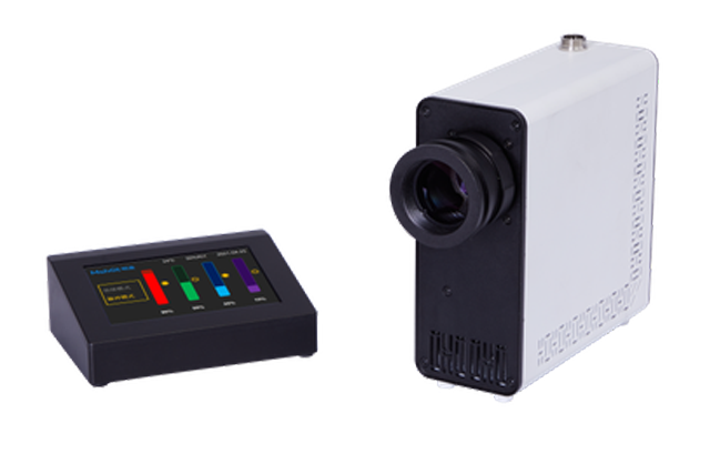
Camera System: The camera system helps record images of the specimen in high resolution. CCD (Charge Coupled Device) cameras are often used in the system. These electron-multiplying cameras excel at imaging single-photon events without losing sensitivity. They require no image intensifier and can capture images at high speeds.
Principles of Fluorescence Microscopy
In fluorescence microscopy, fluorescence refers to a photoluminescence phenomenon: a physical phenomenon in which certain molecules absorb light of a specific wavelength (excitation light) and then emit light of other wavelengths (emission light). This kind of molecule is called a fluorophore. There is a difference between the excitation spectrum and the emission spectrum of the fluorophore. The emitted light energy is lower than the emitted light and the wavelength is longer than the excitation light. This difference is called the “Stokes shift”.
There are two types of fluorescence phenomena: autofluorescence and secondary fluorescence. Autofluorescence means that the sample has its own fluorophore, which can emit fluorescence after being irradiated, such as chloroplasts; secondary fluorescence is also called secondary fluorescence. The sample cannot emit fluorescence after being irradiated. It needs to be labeled with a fluorescent dye first and then irradiated. Fluorescence occurs. Fluorescence microscopy mainly uses secondary fluorescence.
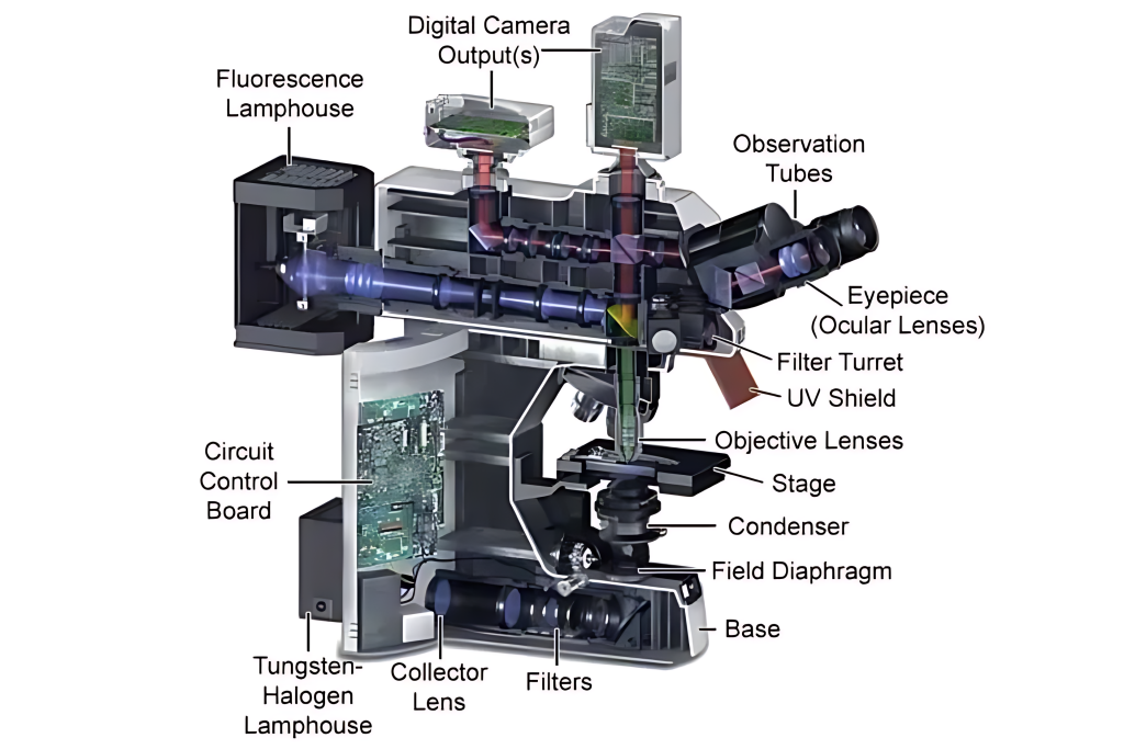
Fluorescence imaging consists of two processes: excitation and emission, which are embodied in the following key components in a fluorescence microscope:
(1) Excitation light source: Provides excitation light with two main indicators
Ø Spectral coverage: A wider spectrum is required to cover the emission spectrum. Different fluorophores require different wavelengths of excitation light, so the excitation light source requires a wider spectrum to cover the needs of various wavelengths of excitation light. If the application scenario is limited to fungal examination, a narrow-spectrum single-channel excitation light source may also be used, but this type of microscope cannot meet the needs of research and other applications.
Ø Emitted light intensity: The light intensity of the light source is different in different wavelength bands. Ideally, the band with a high intensity of emitted light should match the excitation peak of the fluorescent dye. Higher brightness excitation light can produce stronger fluorescence emission.
Nowadays, LED fluorescent light sources with a wide spectrum and high light intensity are mainly used.
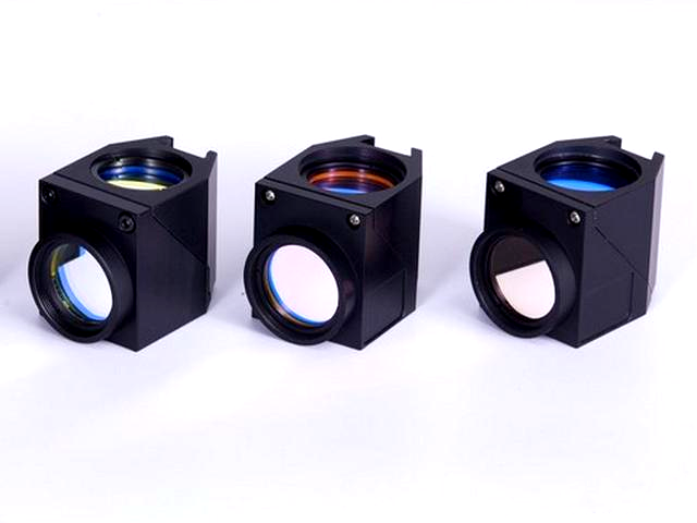
(2) Excitation block: a filter set that forms specific excitation/emission, consisting of three components in a set-up fluorescence microscope
Ø Excitation filter EX: only allows specific wavelength excitation light to pass through to excite specific fluorescent dyes
Ø Emission filter EM: Matches the characteristics of fluorescent dyes and only allows the emitted fluorescence of a specific wavelength to pass through
Ø Dichroic mirror DM: reflects the light in the excitation band and transmits the light in the emission band. This is a component that is available in most current mainstream epi-fluorescence microscopes. Transmission fluorescence microscopes do not have this filter.
The light source in the fluorescence microscope
Fluorescence microscopes use near-monochromatic illumination. There are four common broad sources: xenon arc lamps or mercury vapor lamps with excitation filters, supercontinuum light sources, high-power LEDs or lasers. Lasers are a common choice for confocal microscopy, total internal reflection fluorescence microscopy, and other complex techniques. When using a wide-field epifluorescence microscope, a xenon lamp, a mercury lamp, or an LED with a dichroic excitation filter is generally more suitable. Two microlens arrays in the illumination path of a wide-field epifluorescence microscope can produce an illumination variation coefficient of 1% to 2%, with high uniformity.
Fluorescent sample preparation
Samples suitable for fluorescence microscopy must be fluorescent to be suitable for fluorescence microscopy. Common methods for preparing fluorescent samples include labeling with fluorescent dyes, expressing fluorescent proteins, or utilizing the autofluorescence of the sample.
biofluorescent dyes
Some stains are available for a variety of biological substances. Some small molecules have their own fluorescence and biomolecules bound to them.
Immunofluorescence and fluorescent proteins
The binding of antibodies to their corresponding antigens, thereby labeling proteins or other molecules within cells, is accomplished through the process of immunofluorescence. Modern genetic knowledge and technology allow scientists to edit DNA. The same technique can be used to genetically modify proteins to add fluorescent protein reporters, meaning scientists can make a protein fluoresce. Once it fluoresces, it can be tracked directly, even in living cells.
What is a fluorescence microscope used for?
Fluorescence microscopy is used in biology, biomedicine and materials science. Fluorescence microscopy facilitates accurate and detailed identification of cells and submicroscopic cellular components.
Fluorescence microscopy is also widely used in the field of histochemistry to detect particles that cannot be seen with conventional microscopy, such as the neurotransmitter amines.
It is used in food chemistry to evaluate the presence, structural organization and spatial distribution of specific food ingredients in a product.
There is also a fluorescence speckle microscope. It is a technique that uses fluorescently labeled macromolecular assemblies, such as cytoskeletal proteins, to study movement and turnover rates.
Fluorescence microscopy stains are also used in the field of mineralogy. It is commonly used to study minerals such as coal and graphene oxide.
It is also widely used in the textile industry to analyze fiber dimensions. Epifluorescence microscopy helps study fiber-based materials, including paper and textiles.
Not only that, the use of fluorescence microscopes can also be used to study ceramic porosity with fluorescent dyes and in the field of semiconductor research.