What is differential interference contrast microscopy?
Differential interference contrast microscopy (DIC), also known as Nomarski interference contrast microscopy or Nomarski microscope, is an optical microscope that enhances contrast to observe unstained transparent samples. The image presents a relief-like three-dimensional sense, and It has certain advantages that phase contrast microscopy cannot achieve, and the observation effect is more realistic.
In 1952, Nomarski invented the Differential Interference Contrast Microscope (DIC Microscope) based on the principles of phase contrast microscopy. DIC microscope is also called Nomarski Contrast Microscope. Its advantage is that it can display three-dimensional projection images of structures. Compared with phase contrast microscopy, the specimen can be slightly thicker and the refractive index difference is larger, so the image has a stronger three-dimensional effect.
How differential interference contrast microscopy works
DIC microscopy uses polarizers and birefringent Wollaston or Nomarski prisms to separate the light source into two orthogonally polarized light. The objective focuses these two beams of polarized light onto the sample surface, which is displaced a distance equal to the resolution limit of the microscope. After being collimated by a collimating lens, another Wollaston prism is used to recombine the two polarized lights. The combined light then passes through a second polarizer (also called an analyzer) whose polarization direction is perpendicular to the first polarizer. Interference due to differences in the optical path lengths of the two polarized light beams results in changes in visible brightness. The working principle of a typical DIC microscope is shown in Figure 1:
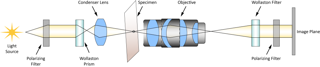
Figure 1: How a typical DIC microscope works. One limitation of DIC microscopy is the increased cost compared to other microscopy techniques. Wollaston prisms, used to separate and recombine different polarization states, are more expensive than the components required for microscopy, such as phase contrast or Hoffman modulation contrast microscopy.
Advantages of Differential Interference Contrast Microscopy
DIC microscopy makes the structure of cells, especially some larger organelles, such as nuclei, mitochondria, etc., have a particularly strong three-dimensional sense, making it suitable for micromanipulation. At present, microscopic operations such as gene injection, nuclear transplantation, transgene, etc. are often performed under this microscope.
Application fields of differential interference contrast microscopy
DIC microscopes are widely used in biomedicine, materials science and other fields. DIC microscopy can be used for high-sensitivity defect detection in transmission materials, especially for identifying laser damage in optical coatings and surfaces. DIC microscopy improves contrast by converting gradients in optical path lengths from changes in refractive index, surface slope, or thickness into intensity differences across a plane. Image slopes, valleys, and surface discontinuities with improved contrast to reveal the contours of surfaces. DIC images appear as three-dimensional undulations corresponding to changes in the optical path length of the sample. Note that the appearance of this 3D model should not be interpreted as an actual 3D model of the sample.
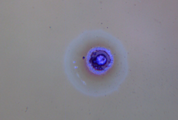
Figure 2: Using a DIC microscope to observe laser damage. Using a DIC microscope can make the slight height difference on the surface of the object produce an obvious relief effect, greatly improving the contrast of the image.
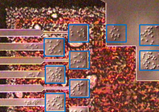
Figure 3: Using a DIC microscope to observe the relief effect of small height differences on the surface of an object